Autism spectrum disorder (ASD) is a neurodevelopmental disorder characterized by neurobiological abnormalities, impaired social skills, repetitive behaviors, and difficulty empathizing with the feelings of others. There is no definitive neurobiological difference that is an indication of ASD, but many areas of the brain have been identified to contribute to the symptoms of ASD.
Cerebral Cortex
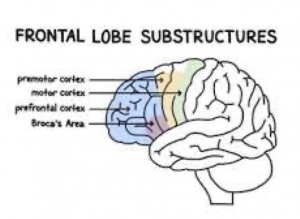
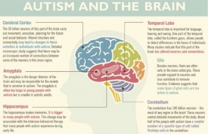
Those with ASD tend to have an overgrowth in the cerebral cortex. This increased volume has effects on the geometry of the brain. All structures in the brain can be affected by an enlarged cerebral cortex because they will have to move to accommodate for the increased volume. Those with ASD can have abnormalities in the folding patterns of the cortical area. Studies have found that the gyri of the  frontal cortex in children and adolescents with ASD are significantly enlarged. The frontal cortex is responsible for managing attention, understanding the feelings of other people, speech and language production, and forming one’s personality. Abnormalities in the frontal cortex associated provide one explanation for the symptoms that are characteristics of ASD.
Cerebellum
The cerebellum of those with ASD has a reduced number of Purkinje cells. Post mortem studies of autistic brains also show an increase in glial cells, reductions of cerebellar nuclei and overall mass, as well as an active inflammatory process within the cerebellum. The decreased number of nuclei within the cerebellum leads to abnormal connectivity with other key areas of the brain. Cognitive deficits, difficulty planning, and impaired working memory are observed in subjects with developmental decreases in the volume of their cerebellum. This video provides interesting information regarding the loss of Purkinje cells and cognitive impairments that are characteristic of ASD.
The cerebellum is also important for fine motor movements, coordination, and balance. Infants with ASD develop basic motor skills later in life than a neurotypical child. Those with ASD often have repetitive hand movements, impaired gait, and slow manual dexterity. The loss of Purkinje cells is one cause of the motor deficits associated with ASD. The cerebellum sends signals to the prefrontal cortex and cortical motor regions; a loss of cells in the cerebellum leads to a loss of connectivity, signal transduction, and activation in key brain regions.
Hippocampus
Those with ASD tend to have a larger hippocampus than those who are neurotypical. A study found that young male children with ASD had a 9% larger hippocampal mass compared to the control subjects. Researchers hypothesized this is due to an increased density of interneurons. Although this hippocampus is typically enlarged in those with ASD, another study found that there is a loss of GABAergic neurons in this area. En2 is a gene linked with ASD and mice with this gene knocked out display typical behaviors associated with ASD. Researchers hypothesized that mice with a double En2 (En2-/-) knockout would have defective GABAergic innervation in the hippocampus.
Results from this study support the researchers’ hypothesis. Mice with the En2-/- phenotype had a reduced number of GABAergic mRNA markers in the hippocampus and cerebral cortex. Abnormalities of the GABAergic neurons within the hippocampus can lead to an unbalanced excitation/inhibition ratio. Loss of these neurons has been shown to lead to impaired maturation of the cerebral and visual cortex. Dysfunction in one area of the brain is rarely isolated to just that area: the brain is the communication center of the body and any deviation from a homeostatic environment can lead to impairments throughout the entirety of the brain.

