Autism Spectrum Disorder (ASD) is a common neurodevelopmental disorder that is diagnosed based on behavioral symptoms. Given the wide-ranging manifestations of the symptoms of ASD, it would be helpful to be able to find more precise diagnostic criteria to provide more specific diagnoses which could allow for more precise treatments. The symptoms of ASD are broadly described as deficits in social communication and interaction as well as restrictive, repetitive behaviors [1]. Since these symptoms are very broad, it is no wonder that there is not thought to be a single cause of ASD. Alternatively, many assorted mechanisms and genetic variations are thought to be associated with behavioral expressions that can be characterized as ASD [2].
The high heritability of ASD is strong evidence for the genetic influence on the variety of manifestations of the condition. This is also supported by the discovery of many de novosingle nucleotide variants as well as transmitted and de novocopy number variants. Because of the wide range of possible contributions and outcomes related to ASD, it is suggested that ASD diagnoses should be subcategorized based on mechanistic commonalities found within different expressions of ASD. One mechanism that could be used to subcategorize ASD is that of dopamine signaling dysfunction [2].
Dopamine Dysfunction in ASD
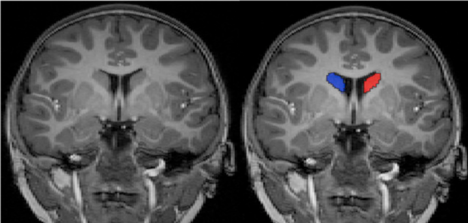
Image 1: The caudate nucleus is a major dopamine hub within the brain. In patients with ASD, this region becomes significantly enlarged, which is shown highlighted in red and blue. [5]A strong piece of evidence for this theory is found in MRI studies of the caudate nucleus. The caudate nucleus is a major target of dopamine signaling. Multiple studies have found enlargement of the caudate nucleus (Image 1) in individuals with ASD. This suggests that ASD is associated with alterations in dopamine related structures and the function of these structures. Similarly, increases in connectivity of the fronto-striatal networks has been positively associated with the severity of repetitive behaviors in ASD. This also links ASD symptoms with abnormal dopaminergic connectivity to cortical structures [2].
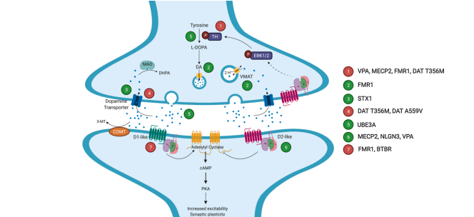
Figure 1: Representation of the dopamine (DA) signaling pathway and ASD associated variants. Red numbers indicate downregulation, reduction, or inhibition. Green numbers indicate upregulation or potentiation [2].There have been many ASD-associated variants within the dopamine signaling pathway reported from various models (Figure 1). Although the direct application of these variants and the degrees of their effects cannot be completely deduced at this time, the significance in this information is that there appears to be many sites of action within the dopamine pathway that are altered in ASD models. Both upregulation and downregulation at specific points within the pathway emphasize the role of dopamine dysfunction in the behavioral manifestations of ASD [2].
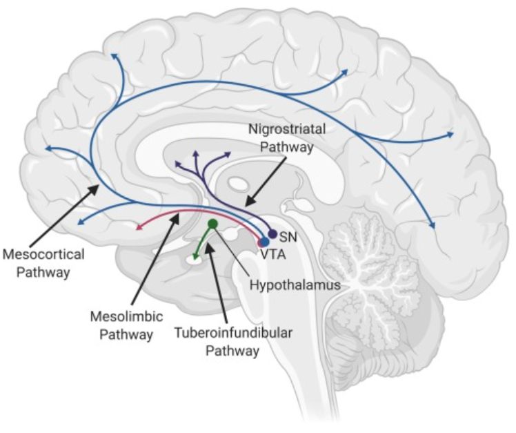
Figure 2: Dopaminergic projection patterns within the brain. Synthesis primarily occurs in the substantia nigra (SN), the ventral tegmental area (VTA), and the hypothalamus. The arrows represent the projections from the sites of synthesis. [2]Given the vast projection patterns of dopamine in the brain (Figure 2), the many variants in dopamine signaling with ASD (Figure 1), as well as the studies linking ASD with changes in dopamine signals and structures, it is difficult to deny the evidence supporting a connection between dysfunction of the dopamine system and ASD behaviors. Dopamine has extensive effects on human behavior including social drive, reward-associated behaviors, allocation of physical energy, attention, and working memory, just to name a few. This broad scope of dopamine related activity could have many implications on the broad scope of ASD manifestations [2].
(2) DiCarlo, G. E.; Wallace, M. T. Modeling Dopamine Dysfunction in Autism Spectrum Disorder: From Invertebrates to Vertebrates. Neuroscience & Biobehavioral Reviews2022, 133, 104494. https://doi.org/10.1016/j.neubiorev.2021.12.017.
(5) Qiu, T.; Chang, C.; Li, Y.; Qian, L.; Xiao, C. Y.; Xiao, T.; Xiao, X.; Xiao, Y. H.; Chu, K. K.; Lewis, M. H.; Ke, X. Two Years Changes in the Development of Caudate Nucleus Are Involved in Restricted Repetitive Behaviors in 2–5-Year-Old Children with Autism Spectrum Disorder. Developmental Cognitive Neuroscience2016, 19, 137–143. https://doi.org/10.1016/j.dcn.2016.02.010.