
Post Traumatic Stress Disorder
The National Institute of Mental Health define PTSD as, “a disorder that develops in some people who have experienced a shocking, scary, or dangerous event.” People affected by this disorder range from war victims to natural disaster survivors. Individuals have experienced the symptoms of PTSD for hundreds of years but it was not recognized until 1980. Names that were used prior to this recognization are “shell shock,” “battle fatigue,” and “soldier’s heart.” DSM-V has now criteria for diagnosis and symptoms for PTSD.
DSM-V:
- At least on stressor such as experiencing the traumatic event (Criteria A)
- At least one re-experiencing symptom such as flashbacks or bad dreams (Criteria B)
- At least one avoidance symptom (Criteria C)
- At least two cognition and mood symptoms (Criteria D)
- At least two arousal and reactivity symptoms (Criteria E)
All of these have to be experienced for at least a month.
Changes in the Brain
The impact of PTSD of an individual varies from the bodies response to the event and the alteration of the structural size or activity of different parts of the brain.
Fight-or-flight is something that can be seen in reaction to a battle or a natural disaster that are known traumatic events. The classic fight-or-flight response that perceives something as a threat is a natural reflex that occurs. The fight-or-flight response is crucial for survival and is important to be regulated correctly. A chronic dysregulation of this can be seen in patients with PTSD.
Dopamine and norepinephrine levels are increased that influence fear conditioning/memories. Glutamate, an excitatory neurotransmitter, increases, while GABA activity decreases. In the brain some structures are altered such as the hippocampus, amygdala, and cortex.
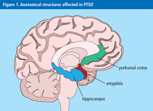
Hippocampus
- The hippocampus is important for the control of stress response and declarative memory. Because the hippocampus has high plasticity an event is able to alter this part of the brain.
- MRI’s showed that in multiple PTSD patients had small hippocampus volumes. The activity found in the hippocampus was lower than normal. The increased levels of glucocorticoid levels and the repeated exposure to this show the toxic effects that this can have on the hippocampus.
Amygdala
- The amygdala is very important in emotional processing and regulating the behavioral responses to events. It is found to be a good mediator for stress responses and emotional learning.
- PTSD patients were seen to have hyperactivity in this brain region when experiencing stressful cues or presentation reminders of the trauma.The impairment discrimination of threat is linked to the hyperactivity of the amygdala.
- Increased amygdala reactivity may be a biological risk factor that increases an individual’s risk of PTSD.
Cortex
- Prefrontal cortex (PFC) exerts inhibitory control of fear responses. PFC is important for things like complex behaviors, including planning, and greatly contributes to personality development.
- A decrease of volume in the frontal cortex was seen. A functional imaging had also shown decreased activation in the PFC when responding to traumatic cues.
- Decreased medial prefrontal activation, reduced of the prefrontal and anterior cingulate volume were observed.
- This impairs the extinction of important fear responses.
References:
- http://pnpcenter.com/index.php/disorders/ptsd-post-traumatic-stress-disorder ▲
- https://www.ncbi.nlm.nih.gov/pmc/articles/PMC3182008/