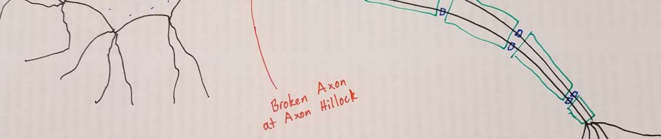
You have probably seen the muscle tremors that affect Parkinson’s patients. Parkinson’s is a disease that kills dopamine-producing neurons in the brain. While traits like intelligence, personality, and memory can decline, the most damage happens to the motor neurons that move our muscles. Over a period of decades, muscle control gradually declines in Parkinson’s patients. There are only a few treatments available for Parkinson’s, and they can only slow the progression of the disease or deal with the symptoms.

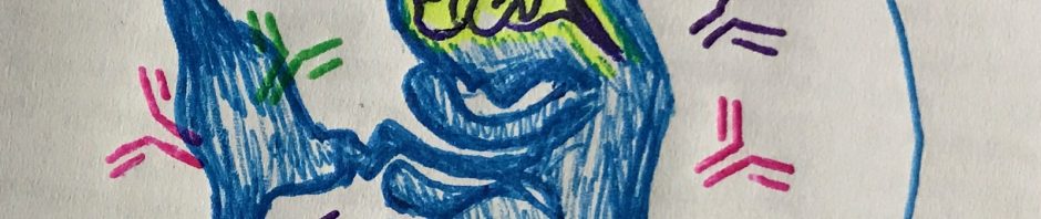
So what is actually killing the motor neurons? The cause is still not completely clear, but we know that Lewy Bodies affect the neurons. Lewy Bodies are clumps of sticky protein that build up in the neurons of people with Parkinson’s. Lewy Bodies are also found in other forms of dementia. They are made up of a protein called alpha-synuclein.
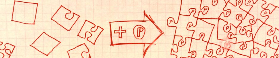
And what makes this protein stick together? The answer might be phosphate groups. Phosphate groups are a molecule of phosphoric acid that gets added onto the protein. For some reason, phosphate groups seem to make alpha-synuclein stick together. Maybe it helps the proteins fit together like puzzle pieces. You probably know phosphoric acid from the label on a can of pop. Drinking phosphoric acid in soda is not dangerous (except to your teeth) because it takes special enzymes to add the phosphate groups onto proteins.
Proteins that tack phosphate groups onto other proteins are known as kinases. It turns out that most kinases are controlled by other kinases, so there is a complicated web of on and off switches inside our neurons. If one kinase is mutated, it can affect dozens of other types of kinases, making them sluggish or hyperactive. This can eventually lead to the clumps of neuron-killing protein.
Interestingly, when one piece of a-synuclein gets a phosphate group it encourages other ones to do the same. This makes alpha-synuclein act like a prion disease in Parkinson’s patients. The prion disease of mad cow disease made headlines when the public learned that their beef cows were eating the brains of other cows. That’s a great way to spread a prion disease, and fortunately humans don’t normally swap much brain tissue. But this does explain how muscle problems can start in an arm or leg and spread to the rest of the body, and how dementia can appear in late stages of the disease. Parkinson’s can start in one area of the brain and slowly march across the brain from neuron to neuron.
The tremors of Parkinson’s make it impossible to eat and difficult to sleep. People that work with their hands for a living are affected even more. If we can stop proteins from sticking together in their neurons, we might be able to stop the disease. Unfortunately, the root cause of the phosphorylation of a-synuclein does not just involve one gene. It will take a lot of work by scientists and probably some supercomputers to put the puzzle pieces together so we can take other puzzle pieces apart.
Concussion Background and Visual Imaging
In the good old days as some people would call it, if you get hit in football or any sport and you were knocked out it was almost seen as a sign of weakness. No man went to the doctor to see what was actually going on in the brain, instead it was recommended to just “shake it off”. However, with the bombshell report that the National Football League was hiding research about concussions and the potential lifelong effects that could result from multiple concussions, the research has become abundant.

Before discussing the research it is important to see the scope of how many concussions actually occur in the United States every year. According to the Centers for Disease Control and Prevention (CDC) in 2013 about 2.8 million Americans were diagnosed with a Traumatic Brain Injury (TBI), most of them being mild TBI’s or as they are commonly known as concussions. These TBI’s accounted for 50,000 deaths in the United States alone. Among all the concussion diagnosed every year it is estimated that 70,000 people experience permanent damage to their brains. One interesting fact, is that the most common sport that causes concussions is not football, it is actually cycling. However, football still experiences just over 36,000 annually.
As a result of a concussion many problems can arise. Cytoskeletal damage can arise because the force of a concussion can cause the complex microstructural components formed by neurons and glia to be damaged. This damage to the cytoskeletal complex can also cause axon dysfunction. This means that the axons on neurons can be overstretched causing them to become overly porous to excitatory molecules. To the extreme end of this the axons can actually be snapped off at the axon hillock. This is severe because this prevents axons from sending action potentials and communicating with the rest of the brain. This can cause cognitive deficits and a lack of communication in the brain. One other main contributor the symptoms of a concussion is increased inflammation. The upregulation of cytokines and cytokine genes causes an increase in stress on neurons. This increased stress if it is prolonged can ultimately lead to cell death and an overall loss of neurons. Either way a concussion can be catastrophic, however what has arisen over the past several decades is the ability to see the effects of a concussion.
This is where the imaging arises. The first form of brain imaging is Diffusion tensor MRI (DT-MRI). It works by sensitizing the MRI signal to the random molecular motion of water molecules (diffusion) by addition of ‘diffusion encoding gradients’ to a standard MR pulse sequences. Water molecules are more likely to travel down white matter (neuronal pathways) than they are to leave these pathways. This allows you to visualize if any of these tracts are damaged, hence seeing if there is a concussion
The second method for visualizing TBI’s is functional magnetic resonance imaging or functional MRI (fMRI). This works by using MRI technology that measures brain activity by detecting changes associated with blood flow. This technique relies on the fact that cerebral blood flow and neuronal activation are coupled. When an area of the brain is in use, blood flow to that region also increases. So in essence decreased blood flow to a region of the brain would imply that there is damage to this area of the brain.
Based on the images above it is clear that concussions are not good for the brain and should be take very seriously. If you or someone you know has a concussion make sure to see a doctor to plot a course of action and an effective treatment.
The Capstone Course Serves as a Sturdy Foundation for the Future
No project is complete without the finishing touch. A Christmas tree lacks luster without the star on top. A painting doesn’t shine without the varnish finish. A Concordia education is incomplete without a capstone course. As a chemistry major, the logical choice for my capstone class was neurochemistry, and I feel it was the perfect finishing touch to my Concordia career.
When I began my freshman year, I was set to be a chemistry major. I was good at chemistry and learning in lecture came easy. Neurochemistry was the first class I took that focused on independent learning, and it happened to be in my last semester at Concordia. I was initially nervous about the amount of biology at the beginning of the course but once we delved into the article analysis, it didn’t seem to matter as much.
While I was doing research at the University of Minnesota this past summer, the grad group I was part of had weekly “Lit Club” where they read current literature published that was similar to their research. I would sit through the meetings every Friday morning thinking that I would never be able to understand the articles they were reading. In neurochemistry, the articles were about the brain and signaling pathways of which I had no background knowledge. I learned that even though I lacked background knowledge, I was still able to understand the general message of each article on my own. It gave me hope that when I am part of a grad group in grad school, I will be able to understand the articles on my own and be able to contribute to the group discussion.
However, this is not the only reason this course has prepared me for the future. It has also taught me to enjoy reading articles of current research findings. Reading articles for class has seemed like a chore in the past, but for neurochemistry, I looked forward to reading about diseases and how altered brain functions resulted in signs and symptoms. I can apply this new enjoyment to my future by continually learning about new scientific discoveries.
As my time at Concordia comes to a close, I have looked back at everything I have learned along the way. Not only have I learned necessary chemistry knowledge, but I have now also learned how to apply my knowledge and continue learning on my own.
I truly believe that this capstone course was the perfect finishing touch on my Concordia education. Learning how to continue learning on your own, for scientists at least, I think is crucial to being a successful scientist. I look forward to my future and learning even more about subjects that I love.
Are Stress and Anxiety Hijacking Your Body?
Stress and Anxiety. Two words you hear often now a days. Especially if you are like me a college student. These two factors can negatively impact our lives and make life much more difficult. These two things are caused by our non-stop desire to be productive. People vary between how they deal with stress and anxiety differently and people differ in how easy they get stressed out or anxious. It’s weird how that is a widely accepted truth. But how does this occur? There are a couple of different reasons why.
A review article in the frontiers of psychiatry reveals that it is the formation of memories attributed to stress and anxiety. Although our bodies are designed in a way that you can make strong memories of stressful events so that in the future you can avoid them. When I say stressful event, the most likely stressor that a human would activate this mechanism would be a wild animal attacking them in the woods or something along the lines of that. But with the modern civilization and the way society is today we do not experience these sort of stressors often at all. This biological mechanism in the brain gets hijacked by the “stressors” we experience as humans almost everyday in the modern world. The paper describes that the adrenal gland releases corticotrophins, which are responsible for releasing adrenaline in the body and increase heart rate and alertness, which is characterized with stress and anxiety. In people with stress and anxiety disorders this becomes dysregulated and this very mechanisms attributes to the formation of the memories associated with the stressors.

In combination with increased corticotrophin release glutamate signaling pays into factor within stress related memories as well. Cellular mechanisms such as a dual histone activation of a protein complex called H3S10P-K14ac increases the production of proteins called c-Fos and Egr-1. These proteins are directly involved with forming strong memories and consolidation processes as well. This underlying mechanism can thus cause an increase of “noisy” thoughts that are characterized with the intense memories of the stressors, which can cause attention deficits. The Prefrontal cortex gets dysregulated as a result of this cascade because the connections between the hippocampus and the amygdala become desensitized.
There is a wide array of medications for anxiety, they include, Selective Serotonin Reuptake Inhibitors (SSRIs), Serotonin-Norephenipherine Reuptake Inhibitors (SNRIs), and Benzodiazepines. The mechanisms for SSRIs and SNRIs block the reuptake channels on neurons of these specific neurotransmitters that play important roles in mood stabilization. Recently it has been described that Histone modification of the phosphorylation and acetylation of the histone H3 complex (H3S10p-K14ac), leads the activation of gene transcription of IEG genes, which facilitate strong memory formation. This mechanism is strongly linked to anxiety disorders and it appears Lorezapam and well-known benzodiazepine blocks the phosphorylation and acetylation of the histone complex. Other anxiety medications and their mechanisms relating to these histone modifications are not well known. However, It has been found that the mechanism for benzodiazepines like lorezapram acts on GABAergic neurons. This relationship could possibly lead to the inhibition of the phosphorylation and acetylation of the histone complex. They are allosteric modulators of the GABA-A receptors. These receptors ligand-gated chloride channel and when activated hyperpolarize the target cell allowing Calcium ions to flow in. Benzodiazepines bind to these receptors in a specific pocket that induces a conformational change and allows GABA to bind.
It’s a terrible cycle. Stress and anxiety when in high doses can be maladaptive for everyone as indicated by this article. However, in this day and age these things are unavoidable. This is why developing a deeper understanding of these aspects and what they can do to a person is important. It is also important to manage these things so that the stress and anxiety levels to become too high where you can develop problems that can be very hard to deal with.
Understanding Concussions and Traumatic Brain Injury
If you were to ask a high school freshman what a concussion is, do you think they would be able to tell you? All I would’ve been able to tell you when I was a high school freshman is that a concussion is when you hit your head really hard. I had no idea really what the consequences of a concussion were and obviously I had a really naïve sense of what it actually is. Luckily that understanding has left me. But I ask….do you think other high school freshmen are different than I was? Sophomores? Juniors? Seniors? What about beyond that? Well hopefully this article can help shed light on what a concussion is and how it can affect your brain after the fact.

A concussion is a traumatic brain injury (TBI) that is characterized by an injury to the head or the body that causes the brain to shake inside of the skull. (Freshman me was partially correct.) If the concussion is severe enough it can cause you to lose consciousness but generally symptoms of a concussion include confusion, slurred speech, sensitivity to light and noise, sluggishness etc. The causes for a concussion are the injuries that result from physical activity such as sports. The most common and famous sport when talking about concussions is football and many of us are aware of the controversy that surrounds it. But why would this be controversial? Well the repeated amount of concussions from football, boxing whatever the activity may be can cause Chronic traumatic encephalopathy (CTE) which is basically a neurodegenerative disorder that is a continuation from repeated concussions that can permanently cause brain damage and worsen the symptoms and premature death. There are an estimated 2-4 million concussions per year in the United States. An article in the Neurosurgery journal discusses the biochemical processes resulting from a traumatic brain injury. It all starts with a high influx of ions into the area affected. This results in overactive cellular responses that kick energy metabolism into overdrive. This can cause damage to the cytoskeleton of the cell and affect the axons that are responsible moving information between cells and within cells. This than can have altered neurotransmission where information is processed more slowly or at an abnormal rate due to the altered neurotransmission. Microglia and supporting neurons will be caused to be overactive and they will release cytokines and trophins that will increase brain inflammation. After all of these sequential events occur the cell or cells will die resulting in neurodegeneration. It is no secret that having repetitive traumatic brain injuries could cause severe brain damage.

This information I believe would be very beneficial to know for people who are involved with sports activities. Parents and players often do not understand the severity of concussions, which makes life difficult for care providers. People need to realize that these symptoms will go away eventually but proper treatment is vital because the risk of severe damage increases during the recovery period of an already in place concussion. This means time for rest and no stimulating activity including intense cognitive thought, which is conducive with schoolwork. Many different factors come into when dealing with a concussion. Teachers, parents, coaches, players etc. all need to come to a realization and a general consensus that concussions are not a topic to throw under the table. Sports and physical activity will not just go away, which means concussions, will not either. However a deeper understanding of what concussions are and how we can treat them
Is Alzheimer’s Disease (AD) Type III Diabetes?
Alzheimer’s disease (AD) is a disease associated with the destruction of brain cells resulting in the decline of cognitive functioning, memory, and social skills. The most common cause of AD is aging. There are approximately 36 million people worldwide who suffer from Alzheimer’s. Since AD is common and not entirely understood, it is important to better understand the processes regulating AD progression in order to develop more targeted treatments and interventions that can delay or prevent the disease.

Something I didn’t realize about Alzheimer’s disease (AD), until we talked about it in our Neurochemistry capstone class this week is that insulin has a compelling role in the progression of the disease. Obviously AD is multifactorial, since things like hyperphosphorylated tau and AB plaques are other problematic contributors to the disease. But interestingly, several studies have shown that overactivation of the P13-k/Akt/ mTOR signaling pathway is an early feature of AD.
Insulin is one of the key activators of this P13-k/Akt/ mTOR signaling pathway. While signaling through this pathway, it is crucial for the cell to maintain the proper activity level. Proper signaling through the P13-k/Akt/ mTOR signaling pathway allows for the beneficial processes resulting in DNA repair, cellular repair, translation of anti-stress proteins, and overall maintenance of healthy cell systems within the neurons of the brain.
Evidence is suggesting that insulin resistance is problematic occurrence within the neurons of people with AD. Insulin resistance does not allow the proper regulation of the P13-k/Akt/ mTOR signaling pathway because of sustained activation. This is a problem because insulin resistance in AD neurons has been associated with decreases in episodic and working memory, resulting in the symptoms of AD.

So how does this relate to diabetes? Well Type I Diabetes (T1D) is associated with hyperglycemia because the beta cells of the pancreas that produce insulin are destroyed by the person’s own body, resulting in decreased insulin action. Type II Diabetes (T2D) is associated with insulin resistance in peripheral tissues due to low insulin receptor expression and activity. Like T2D, AD can be associated with insulin resistance, but in this case confined to the neurons of the brain. In this way, AD could be considered Type III Diabetes. With this in mind, future research should consider treatments for AD that can minimize the impacts of insulin resistance in the brain.
Maternal Autoimmunity and Its Implications in Autism
Autism spectrum disorder (ASD) is a neurodevelopmental disorder that is known to impair a child’s communication, social behavior, and contribute to the characteristic repetitive behaviors. Like many neurological impairments, ASD has many possible contributing factors to the disorder. In our studies this week during our Neurochemistry course, the paper we discussed outlined prenatal viral infections, zinc deficiency, abnormal melatonin synthesis, maternal diabetes, prenatal stress, toxins, and parental age as possible environmental factors that influence the development of autism.

The risk factor that I found particularly interesting to the story is maternal autoimmunity. This also relates to the age of the mother in children with autism because the majority of autoimmune disorders arise when someone is 30 years of age or older. Autoimmunity is an immune response of the body that attacks and destroys the body itself. A couple of examples of autoimmune disorders are type I diabetes mellitus, myasthenia gravis, and lupus. (Not all types of maternal autoimmunity will result in the development of ASD)
Why an autoimmune disorder is problematic for fetal neural development is that even maternal autoantibodies are able to cross the placental barrier. The placenta normally provides a selective barrier that allows immune factors and molecules for nutrition to transfer to the developing fetus, while restricting the passage of other potentially harmful molecules like pathogens. While it is a positive that the placenta allows the mother to give some passive immunity to the fetus during development, this same process is a negative for the baby when their mothers exhibit autoimmunity. A normal maternal IgG will provide the baby with protective immunity from the mother’s environmental exposures until it is able to have an immune response on its own. Unfortunately, auto reactive IgGs from the mother will also be cross the placental barrier, recognizing self-proteins and possibly interfere with fetal development.

Another interesting fact about fetal development is that the blood brain barrier of the fetus is not fully developed for the majority of gestation. For mothers, their own blood brain barrier is important because it normally restricts auto reactive IgG from the CNS. Since fetal protein reactive IgGs are able to cross the placental barrier during gestation, these IgGs may have access to the developing brain of the fetus that does not have the fully developed blood brain barrier to protect itself. It is therefore possible that fetal protein reactive IgGs of the mother that have access to the fetal brain could interfere with CNS development of the baby and result in neurodevelopmental disorders like autism. Several studies have reported that fetal brain reactive antibodies are more commonly found in the circulation of mothers of children with certain subsets of ASD.
Better understanding of autoimmune disorders and especially fetal brain reactive antibodies will be important for considering the mechanisms that fetal exposure to maternal autoantibodies have in the development of ASD.

Treatments for the Cause or the Symptom?
Parkinson’s disease….. A very tough disease to deal with. It seems the more you know about this disease the less you do actually know. Upon learning about PD I found myself asking more questions than I had to start. Lets see what you think…..
Parkinson’s Disease is a progressive disorder that affects the movement of person suffering with the disease. Around 1 million people in the United states suffer from Parkinson’s Disease and each year, around 60,000 new cases are diagnosed. The disease is characterized as a loss of dopamine neurotransmission in the human brain. This results in all of the symptoms characterized with PD such as, tremors, slowed movement, rigid muscles, impaired posture and balance, speech difficulties etc. These symptoms come at the cost of decreased dopamine neurotransmission that fuels the neurons and connections in your brain.
The current treatments for Parkinson’s disease are mainly to treat the symptoms of the disease. L-Dopa or levodopa is an amino acid that is a precursor to neurotransmitters dopamine, norepinephrine and epinephrine. It is able to cross the blood brain barrier and subsequently be converted into dopamine. Early studies showed that L-DOPA can cause sever side effects such as nausea and vomiting. These side effects were occurring because the L-DOPA was being converted into dopamine before it was crossing the blood brain barrier. So researchers added inhibitors of dopa-decarboxylase, which is the enzyme responsible for converted levodopa into dopamine; these inhibitors are called carbidopa and benserazide. L-DOPA medication provides relief to the tremors and shakiness symptoms of Parkinson’s disease. However, long-term use of the drug is shown to induce dyskinesia for 80% of patients that use levodopa. Dyskinesia is a disorder characterized by involuntary muscle movement. As patients progress with the treatment of levodopa the drug effects begin to wear off several hours of some or all doses of the medication. The reasons these motor fluctuations occur is unclear but is thought to be because of uneven changes in the basal ganglia circuitry resulting in the depletion of both dopamine and the drug treatment. Treatment of levodopa-induced dyskinesia is difficult but treatments used for the disease or dopamine agonists and neurosurgery. The main problems with both Parkinson’s disease and LID are the depletion of dopamine or the lack of dopamine circulating throughout the brain.
So what is the cause for this lack of dopamine Neurotransmission? Why do people get Parkinson’s Disease? Several factors point to genes. Specifically the SNCA gene which encodes for a-synuclein protein. A review article in the frontiers in molecular neuroscience journal discusses this very protein and why it is highly involved in Parkinson’s disease. In a general sense, a-synuclein is being turned on in a specific area on the protein called Serine 129 (S129) which is called hyperphosphorylation. This attributes to the formation of Lewy bodies which consequently, cause the cell to die and thus no dopamine present.

The article also goes into detail of different proteins that are related to the mechanism for the hyperphosphorylation of a-synuclein, such as polo-like kinase, casein kinases, and G protein coupled receptor kinase. However, more research needs to go into this mechanism in order to establish more of an understanding of this phenomenon. In addition to the hyperphosphorylation of a-synuclein, other factors such as the dysredulation of kinase proteins are attributed in the pathogenesis of PD. These kinases include proteins called LRRK2 and PINK1. Like the hyperphsophorylation of a-synuclein these are due to genetic factors and more research needs to be done.
Like I said, this disease is very complicated to understand. It seems like the more you know the less. Hoever, I reall think that this disease needs to be researched more and I believe eventually we can come up with an effective way to treat the causality of the disease not just the symptoms.
Removing Addiction From the Shadows
The American Society of Addiction Medicine describes addiction as a chronic disease of brain reward and motivation systems. Affected individuals do not have the ability to abstain, control, and recognize problems with one’s behavior.
Risk Factors
Environmental factors influence and individuals genetic risk factors which alone account for roughly half of the probability that someone will become addicted during their life time. This combination effect determines how likely addiction is to manifest in any individual. Although, studies have shown that life experiences such as education from parents can infact minimize risk. Other factors can increase risk such as experiencing a trauma, depression, and damaged social connections.

Current Research
Addictive drugs alter transmission within the brain. These drugs of abuse impact the complex signaling pathways involved with reward learning. Specifically, complex dopamine and glutamate signaling pathways are altered by changes to secondary signaling molecules such Calcium and cAMP.
Protein kinase ability is also decrease which are crucial for the phosphorylation and de-phosphorylation mechanisms necessary for proper synaptic plasticity. These drugs also dysregulate complex dopamine and glutamate signaling networks further affecting synaptic plasticity and function. All of which contributes to the progression of a healthy brain to an addicted state.
Harmful Attitudes Towards Addiction
This disease commonly carries negative stereotypes, misconceptions, and harsh judgement that create barriers to public awareness and treatment for affected individuals. Common beliefs persist in our society such that affected individuals lack the desire, willpower, and morals to simply choose to quit.
Addiction is a very complex disease that can happen before an individual realizes what is going on and often necessitates more than just pure will power to overcome. Recently, I even stumbled across a social media article with a direct message to individuals struggling with addiction to stop calling it a disease and labeling it as a ‘pity identity’.

One of the main stereotypes surrounding addiction is that it is a choice. In some cases, in a very basic sense may be true for some cases.
However, humans make mistakes and I do not believe that anyone experiments with a drug to intentionally become addicted. In many cases, it is a unintentional byproduct of medical care, and other environmental factors.
Medical issues such as chronic pain can lead to substance abuse problems. Opioid addiction often begins by taking pain medications given in a medical setting to manage a condition. People assume that medications received from a medical setting are safer to use than an illicit drug. However, individual biological risk factors can lead some individuals down the addiction pathway from something as simple managing back pain. One study found that up 80% of heroin addicts report their first opioid use was in the form of a prescription drug. It is a situational risk that everyone needs to be aware of to make informed decisions regarding their health.
It is a simple fact that certain individuals are just more susceptible than others for developing an addiction. Eluding the mechanisms driving addiction and underlying risk factors are essential for removing stigma, and providing better treatment and prevention.
Type III Diabetes: Alzheimer’s Disease
Many may wonder the rationale behind this title, and indeed it seems a strange connection to make. But the truth of Alzheimer’s disease lies in insulin receptors, and eventually insulin resistance. These receptors are, of course, localized to the brain, and not the pancreas (as in the case of diabetes as we know it). To understand the idea of Alzheimer’s as an insulin disorder, we must initially observe and understand the properties of the receptor on a cell.

Insulin is, at its very simplest, a growth factor. This means, in short, that it promotes cell longevity and proliferation. Growth factors could lead to a plethora of anti-aging medicines. And just so, we can also realize that an enormous risk factor for Alzheimer’s disease is aging. Certainly, if aging is happening, insulin reception is not (or at least not as well as it normally does). This is the first connection of Alzheimer’s with insulin resistance.
But if that were the only connection, it would likely be coincidence. We must delve deeper into the causes of Alzheimer’s, or at least the causes doctors have identified thus far. The beta-amyloid plaque has been used as the sure-sign of Alzheimer’s for decades. This is an aggregation of protein that essentially clogs up neurons and eventually causes their degeneration. But what could these beta-amyloids have to do with insulin resistance? The answer lies in competition.

Many body receptors have a certain ligand/molecule they like to bind to, but nothing is perfect, and other molecules can also bind instead. It has been proposed that beta-amyloid plaques can bind to insulin receptors on neurons and essentially cause them to not recognize when insulin is actually present. This leads to the opposite effect of the insulin “growth factor” and instead causes the cell to degenerate. Even further, since the cell is recognizing that many of these receptors are full of binding molecules, it “decouples” the receptors from the membrane, making insulin signaling even more difficult.
In reverse, studies have indicated that low levels of insulin reception cause levels of beta-amyloid plaques to increase, exacerbating the problem. There is still no consensus on whether low insulin reception initially creates high levels of beta-amyloid, and this feedback causes lower levels of insulin reception, or whether high levels of beta-amyloid begin the process. Either way, this emphasizes the “insulin resistance” that is so common with diabetes.

All of this culminates in the treatment for Alzheimer’s. A newly-prescribed medicine is intranasal insulin spray. By introducing the molecule to the nasal membranes, it quickly makes its way into the brain, increasing insulin levels. These increased levels might be able to “push” the beta-amyloid plaques out of the insulin receptors and allow normal binding and signaling to occur. Whether this is actually the case is unknown, but the medicine shows promising results.
Is this just one more reason to eat a healthy diet and exercise frequently? Perhaps. But in the end, the choice is our own. We must simply realize that our actions today have an effect on us for the rest of our lives.


