My Concussion Experience

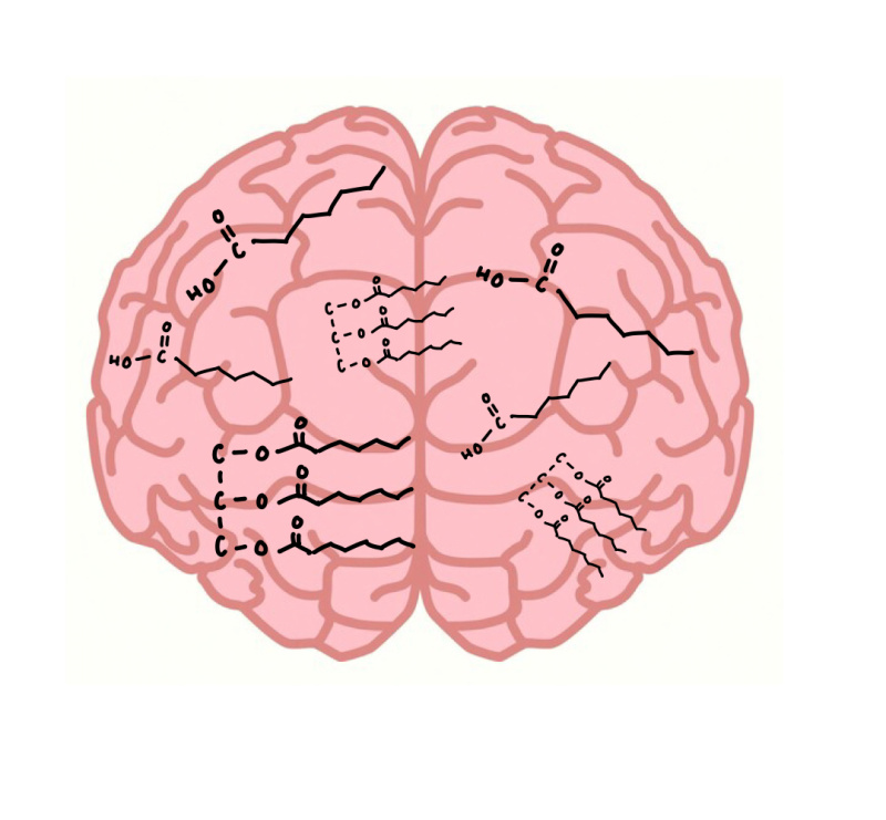
Fig. 1 A visual representation of a concussion. Picture obtained from brainfacts.com.

Story Time:

I have never experienced a concussion, nor have I ever seen someone I’m close to get a concussion. This statement would’ve been true a year ago, but I can no longer say this is the case. This past March, one of my best friends got a concussion and it was an event I will never forget. I was at this friend’s house, hanging out with her roommates and mine while we waited for her to get off of work. She had been snapchatting us throughout the evening, telling us about the drama going down at work. She had told us that she got hit in the head amidst the drama, but she wasn’t feeling anything more than a headache. She arrived at her apartment 20 minutes later, greeting us but excusing herself to her room to lay down. Almost two hours later, my friend came out of her room, tears streaming down her face as she held a hand to her head. She looked at my roommate and I and asked, “when did you guys get here?” Immediately, our concern skyrocketed. We replied that we had been there since nine o’clock, and she had greeted us when she came in the door. She then proceeded to ask, “how did I get home?” My roommate and I looked at each other, mutually deciding that we needed to take our friend to the emergency room. We continued to ask our confused friend questions about what day it was, what she remembered, and how she was feeling as we prepared her to go. We helped her get her coat on and grabbed her ID before taking her down to the car. I gave my friend a blanket that she could use to block out the lights, and we were on our way to the emergency room.

Upon arrival to the emergency room, I knew my friend’s condition had worsened. My friend was squinting at the light as we stepped out of the car. She was very unsteady as we walked to the front doors of the emergency room—I was essentially holding her up, at this point. At this time, COVID was still a severe problem, so they were not allowing people other than the patient into the ER. However, the person at the COVID checkpoint saw the pair of us and said nothing as I walked my friend to registration. I told my friend to close her eyes as I checked her in, as she couldn’t even remember her birth date. I went to the front desk and told her who my friend was, what had happened, and gave her my friend’s photo ID. A nurse came out with a wheelchair just a few minutes later, and the two of us got my friend seated in the wheelchair. The nurse took my friend to the back, while I waited in my car in the parking lot. My roommate and I were waiting for our friend at the emergency room until 3:00 am.

Post-Concussion Thoughts:

Whenever I thought about concussions prior to the incident with my friend, I imagined how much it would hurt to be hit that hard in the head, but I never thought about the actual injury itself. I never thought about the memory loss, the confusion, the photophobia, or the  loss of balance one may experience after the event. It wasn’t until I witnessed it with my friend that I began to think about all of the conditions associated with concussion. Figure 2 shows a more complete list of concussion symptoms, but I will focus on only a few.

Fig. 2. A more extensive list of concussion symptoms. Image obtained from Beth Sissons’ article of “What to Know About Post-Concussion Syndrome” on medicalnewstoday.com

I also have never thought about the biological causes behind these conditions. The migraines, light sensitivity, and sound sensitivity can all be attributed to the ion flux that occurs following a concussion.[1] Ion flux is the movement of ions into and out of the cell. This flux occurs because a concussion makes little holes in the plasma membrane, which allows calcium and sodium to move into the cell and potassium to move out of the cell.1 A good model for this idea is the sharing of a twin bed. If one person is in the bed, and another joins, the original person is likely to leave (unless they fight for it, but let’s imagine this occurs in a perfect utopian society). This movement of ions can cause depolarization of the membrane, leading to excess release of glutamate.1 The impaired cognition, slowed reaction times, and slowed processing often seen post-TBI are attributed to impaired neurotransmission and axonal dysfunction.1 Impaired neurotransmission is an imbalance in the release of excitatory and inhibitory neurotransmitters. There are more excitatory neurotransmitters (action-inducing chemicals) binding to receptors than inhibitory neurotransmitters (action-inhibiting chemicals).1 The axonal injury is the stretching of the axons of neurons.1 Like a hair tie that you’ve used one-too many times, the axon stretches out and weakens as a result of the trauma to the brain. All of these things were going on in my friend’s brain, lasting months after her injury. You don’t really think about any of these severe effects from a concussion until you are a witness to it. I will remember this event forever, and I am glad that I am able to connect my friend’s symptoms to biological events going on within her brain.

[1] https://moodle.cord.edu/pluginfile.php/1052900/mod_resource/content/3/2014%20The_New_Neurometabolic_Cascade_of_Concussion.3.pdf

Obesity: Whose Fault is it Anyway?

Stats

In 2018, 139.7 million US adults were considered obese (CDC, 2021). This is an increase from 30.5% in 2000, to a whopping 42.4% in 2018 (CDC, 2021). To further break down these figures, 49.6% of Black adults, 44.8% of Hispanic adults, 42.2% of White adults, and 17.4% of Asian adults were considered obese in 2018 (CDC, 2021). Obesity prevalence is also correlated with socioeconomic status (SES) and amount of education received (CDC, 2021). Interestingly, men are least obese at the high and low ends of SES while women are least obese in only the highest SES (CDC, 2021). For more information, visit the CDC’s website here.

This high rate of obesity is destructive for many reasons. The most salient issue is directed at the obese themselves. Being obese can drastically increase one’s risk for cancer, heart disease, stroke, and type 2 diabetes (CDC, 2021). Additionally, the medical cost of obesity in the US impacts the obese, employers, and the government (through funding Medicare and Medicaid). The medical cost of obesity is estimated to be between $147 and $210 billion every year (GWU, 2013). So whether it’s for health of economic reasons, we should try to understand the underlying neurochemical cause of obesity.

Overview Of Science

When trying to understand the neurochemistry of obesity, it is helpful to have a guide in front of you. To aid in your understanding, please refer to figure one below while reading the next section.

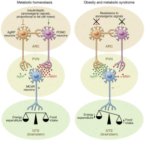
Figure 1: Obesity Pathway Artstract by Bretton Badenoch. The left side (red) shows some of the pathways that lead to dysregulation of hunger. The right side (in blue) demonstrates what neurochemistry looks like in a non-obese brain.First, we will take a look at the right side of figure 1. This side shows how appetite is controlled in a non-obese brain. Two of the most important molecules for controlling appetite are insulin and leptin. After eating a meal, insulin and leptin levels increase throughout the body.  Insulin will then bind to an insulin receptor that ultimately causes an increased activation inproopiomelanocorticin (POMC) neurons and an inhibition of AgRP neurons (Jais, 2017). Leptin will have the same outcome, but acts on a separate leptin receptor (Jais, 2017). The decreased AgRP leads to depressed appetite while increased POMC activation promotes a feeling of fullness (Jais, 2017).

There are multiple ways for this pathway to go awry. Saturated fatty acids (SFA), and TNFalpha (a cytokine caused by inflammation) both contribute to the pathology of obesity through dysregulating hunger (Jais, 2017). The left side of figure one shows how these molecules interfere with POMC and AgRP activation leading to a feeling of hunger (Jais, 2017). This overactive feeling of hunger can lead to overeating and obesity.

Conclusion

Obesity is a disease that affects that affects nearly half of Americans (CDC, 2021). Because of this, it is a very costly disease both to the individual and society at large (GWU, 2013). Now that we know a bit of the neurochemistry underlying obesity, perhaps we can find more empathy for the obese. Obesity is often blamed on the individual for having weak willpower, but the chemistry shifts the blame to something beyond willpower. The connection between willpower and elevated levels of TNFalpha is tenuous at best.

It seems unlikely to me that willpower can decrease levels of inflammatory TNFalpha in any meaningful way. Rather, it’s possessing the knowledge on how to decrease levels of SFA and TNFalpha that seem most potent in preventing and reversing obesity. But that’s a topic for another blog post.

Citations

  1. CDC. (2021, September 30). Adult obesity facts. Centers for Disease Control and Prevention. Retrieved November 21, 2021, from https://www.cdc.gov/obesity/data/adult.html.
  2. George Washington University. (2013). Fast facts: The cost of obesity – George Washington University. Fast Facts Cost of Obesity. Retrieved November 21, 2021, from https://stop.publichealth.gwu.edu/sites/stop.publichealth.gwu.edu/files/documents/Fast%20Facts%20Cost%20of%20Obesity.pdf.
  3. Jais, A., & Brüning JC. (2017). Hypothalamic inflammation in obesity and metabolic disease. The Journal of Clinical Investigation, 127(1), 24–32. https://doi.org/10.1172/JCI88878

The US Obesity Epidemic

Uncle Sam Overweight | The Edible Eighteenth Century

Image 1

The real Captain America is obese and bad at math - CNET

Image 2

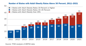
The obesity rate within America during 2017-2018 sat at a staggering 42.4%. If that wasn’t bad enough, 73.6% of Americans are now considered to be overweight. Look at the chart below, obesity rates within the US have increased exponentially since the 60’s. Several drastic changes occurred during the 80’s within American culture/society that impacted obesity rates, leading to the exceptionally high rates of obesity.

Figure 1

Obesity is associated with many negative health conditions. These conditions cost America anywhere between 147 and 210 billion dollars annually. Will our societal eating habits become so greatly problematic to the point of Wall-E? As a kid I thought the movie was funny, the portrayal of the overweight people seemed to be amusing and unrealistic since there appeared to be a complete lack of healthy humans. However, if Americans continues this trend towards increased obesity, we could end up looking similar to those pictured in this fictional movie. Would I still find it funny, probably not.This Is The Hidden Cannibalism You Missed In Wall-E - UNILAD

Picture credits; Wall-E

One of the changes mentioned earlier that took place in the 80’s relates to the change in meal composition. As a society we are moving away from healthier meals in favor of high fat diets. We talked at length about this in class, there are several negative consequences of eating a high fat diet. You can find that information on this document if you would like to look more into it. As an example of how we’ve changed out consumption of different foods, lets look at a couple of examples. Consumption of vegetable oil within Americans diets has DRASTICALLY increased over time. 400 additional calories are consumed daily due to this one ingredient alone. Unfortunately that is not the only food “category” that has changed. Look below to see examples of three food categories and how their average (daily) caloric intake has changed over time.

Figure 2, 3, and 4

Here is a list of the top calorie sources within the US diet

1. Grain-based desserts (like cakes, cookies, and donuts)

2. Breads made with yeast

3. Chicken and chicken dishes

4. Soda and sports drinks

5. Pizza

6. Alcoholic beverages

7. Pasta and pasta dishes

8. Mexican dishes

9. Beef and beef dishes

10. Dairy desserts (like ice cream and cheesecake)

In case you hadn’t noticed, none of these are overly healthy options and are all very high in fat, leading back to the article relating to high fat diets and how unhealthy that is for us.

The American diet in terms of daily calories has changed over time. Today Americans consume more than 3,600 calories every day which is a 24% increase from 1961 when Americans only consumed 2,880 calories daily. The chart below shows how the US food chain has ballooned over time in order to meet the demands of the consumers. The chart shows slightly different estimates of daily caloric intake, estimates that vary per source, but it shows the shift from 2,880 to 3,600 calories. Sources seem to have conflicting estimates, but almost all seem to agree that daily caloric intake has risen by at least 20%.

Figure 5

What changes have lead Americans to become so obese, especially the large spike in obesity during the 80’s? Well the simple answer includes several things: a change in the composition of meals (for example more saturated fat and vegetable oil), source of food (fast food over home cooking), and lifestyle changes (less exercise and more loungin around watchin the Vikings lose).

In terms of a changing food source, the graph below does a good job showing where families have gotten their food from over the years. Families used to eat at home, now fast food and restaurants compose a much larger percentage of weekly meals.

Unfortunately obesity in America is not only a problem of consumption, it is also a problem of physical exertion. What do I mean by this? Well, most Americans fail to meet the US governments minimum daily recommended exercise. That recommendation includes 75 minutes of vigorous cardiovascular exercise a week as well as muscle strengthening twice a week. Sadly only 23% of Americans achieve this minimal standard. Time is spent doing other things, for example watching television.

Figure 6

Figure 7

With Americans having a higher caloric intake and a decrease in exercise it is not shocking to see a drastic rise in obesity. Lets all get out of our chairs, eat a little better, and work as a society to better our health. If we don’t and our diets continue to contain a high ratio of fat, we might get to a point where our body becomes resistant to both insulin and leptin. This scenario would lead to a detrimental feedback loop that would benefit no one, leading to even higher rates of obesity with little hope for individual health and well being.

Here’s a link to a video of me explaining my topic if you’d rather listen to my voice instead of reading: https://drive.google.com/file/d/1W9P1wZxyiVZsQILe2YTxtd4Sm4rKFIo-/view?usp=sharing


Centers for Disease Control and Prevention. (2021, September 30). Adult obesity facts. Centers for Disease Control and Prevention. Retrieved November 16, 2021, from https://www.cdc.gov/obesity/data/adult.html.

Centers for Disease Control and Prevention. (2021, September 10). FastStats – overweight prevalence. Centers for Disease Control and Prevention. Retrieved November 16, 2021, from https://www.cdc.gov/nchs/fastats/obesity-overweight.htm.

Schuler, L. (2021, February 12). The real reason Americans are so fat. Men’s Health. Retrieved November 16, 2021, from https://www.menshealth.com/weight-loss/a19536794/reasons-americans-are-fat/.

” why the world is overweight? the 1980’s and its contribution to our health now! Size Fantastic – Sustainable Weight Control – Healthy Weight Control and Weight Loss. (n.d.). Retrieved November 16, 2021, from https://sizefantastic.com.au/weight-loss-tips/why-the-world-is-overweight-the-1980s-and-its-contribution-to-our-health-now/.


A Brief History of Anxiety: Were Dinosaurs Ever Anxious?

In today’s world we are taught to treat the condition “anxiety” as a potential mental disorder, but this does not mean it has always been that way in history. Before further discussing where the illness came from or when it was first recognized, we must understand what it really is today.

What is Anxiety?

Artstract by Zsofia Zelenak

Anxiety has existed ever since the need of survival, the kind of anxiety that is beneficial to us evolutionarily. This kind of anxiety has a role in the whole fight or flight system and keeps us alive, but that is very different from the anxiety we recognize today. When we talk about anxiety disorders we mean that the general nervousness, fear, and stress are at the extremes in everyday life and interferes with a person’s life. As to what causes anxiety, there is a few ideas out there, such as:

  • imbalance in brain signaling in areas of the amygdala and hippocampus
  • excess glutamate (resulting in the overstimulation of neurons)
  • lack of adequate GABA (resulting in the inhibition of neurons)

Where anxiety came from and where it is now?

Ancient Greece: the very first documented signs of having anxiety described. With the then popular “hysteria” came the description of negative anxiety symptoms.

Early Renaissance: or the witch era. Women who were simply anxious, or again, “hysteric” were often accused of being witches.

Victorians: the trend here took a tiny bit of a different rout, instead of calling them witches they were now considered crazy. It used to be common for family members to take those who had panic attacks and get them treated at an insane asylum. Treatments included electroshock therapy, and other cruel torture rather than the therapies we see today.

The American Civil War: or the time of “soldiers on drugs”. Soldiers who returned from the war were thought to have “irritable heart syndrome”, which is the term used for the condition we now call PTSD.

The 18th Century: “irritable heart syndrome” was now the new “nerve weakness”, which doctors decided to treat with alcohol and bromide salts. It is also important that this was a turning point in anxiety reaching other than women, it was now men that were being targeted for this illness.

The 20th Century: Although it is unclear, but some documents support the idea that Russia was amongst the first to recognize anxiety as being psychological, rather than simply physical. Psychiatrists working with war veterans now began to use barbiturates, not to cure, but only to treat symptoms and sedate these men. Therapies started spreading as a form of treatment option available, but was still rather brutal than helpful.

Late  20th Century: from around the 1950’s, modern medicine began to develop, and options such as electroshock therapy were mostly eliminated and left only for extreme cases. Fear exposure therapy was one that arose from this time, as well as the start of antidepressant use for not only depression, but anxiety. It was not until the 1980’s that the term “anxiety disorder” was created, leading to its official recognition by the American Psychiatric Association. From this time on, researching this disorder became much easier.

As for Today: The DSM-5 (2013) recognized anxiety and describes it as “Excessive anxiety and worry (apprehensive expectation) about a number of events or activities. Difficult to control the worry.”


Sources:

https://www.ncbi.nlm.nih.gov/pmc/articles/PMC5573555/

https://www.ncbi.nlm.nih.gov/pmc/articles/PMC4610616/

https://www.calmclinic.com/brief-history-of-anxiety

Let’s Talk About Dietary Fats

Types of Dietary Fats

Fats are a necessary part of an individual’s diet. Some benefits include organ protection, nutrient absorption, and energy metabolism. There are three main types of dietary fats: unsaturated, saturated, and trans-fat. Saturated fats are solid at room temperature, and are known to increase one’s cholesterol levels, specifically the ‘bad’ cholesterol, LDLs. The most common American foods with a  high saturated fat content include:

  • Pizza and cheese

            Figure 1
  • Whole and reduced fat milk
  • Meat products (beef, sausage, etc.)
  • Grain-based desserts (cookies, brownies, etc.)
  • Variety of fast food

Saturated fat is found mainly in foods from animals, but may also be found in plants such as coconut and palm oil (coconut oil may not be as great as you think)! On the      other hand, we have unsaturated fats. These are liquid at room temperature and are considered to be the ‘good’ fats through improving blood cholesterol levels (lowering LDLs and raising HDLs), easing inflammation, and stabilizing the heart. There are two types of unsaturated fats: monounsaturated and polyunsaturated.

Figure 2
Figure 2
  1. Monounsaturated fats:
    1. Oils (peanut, canola, olive)
    2. Avocado
    3. Nuts (almonds, pecans, peanuts)
    4. Seeds (pumpkin, sesame, etc.)
  2. Polyunsaturated fats:
    1. Oils (sunflower, corn, soybean, flaxseed)
    2. Walnuts
    3. Flaxseeds
    4. Seafood

Within polyunsaturated fats, there are fatty acids known as omega-3. The body is unable to produce omega-3 fats on its known, so it is important to obtain these from one’s diet. These are mainly found in seafood (fish) and walnuts or flaxseeds. Lastly, trans-fats is a fat produced through the process of hydrogenation to elongate shelf-life. This fat is considered to be the most detrimental to one’s health through increased inflammation, insulin resistance, and raise bad LDLs while lowering good HDLs. These are often known as partially hydrogenated oils (PHOs), and are banned in many countries.

Saturated Fatty Acids (SFAs) in the Brain

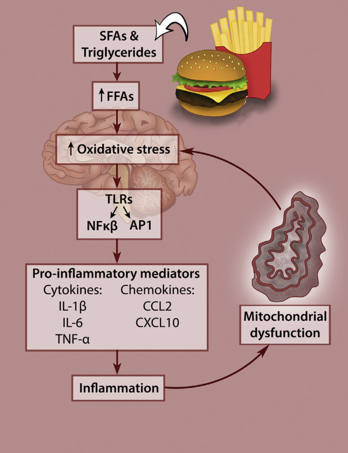
It has been shown that specific fatty acids, specifically saturated fatty acids (SFAs), are linked to hypothalamic inflammation, disrupt insulin and leptin signaling, as well as trigger inflammatory signaling cascades. The induction of SFAs are able to trigger Toll-like receptor (TLR2/4) signaling. TLR4 activates the response gene, MyD88, activates the kinase complex, IKK. Under conditions of high-fat diets (HFDs), IKK phosphorylates IĸBα, an inhibitor of the pro-inflammatory cytokine producer, NF-ĸB. This allows NF-ĸB to translocate to the nucleus of the cell and transcribe pro-inflammatory cytokines such as TNF-α and IL-1ß, as well as SOCS3. This signaling cascade increases neuronal inflammation in brain tissue as SFAs are able to cross the blood-brain-barrier (BBB) (Jais and Brüning, 2017). SFAs also disrupt insulin signaling through the increased production of ceramides within the brain. Ceramides activate protein kinase C (PKC), which is a primary inhibitor of PI3K. PI3K plays a major role in the insulin signaling pathway, and its inhibition will cause nuclear exclusion of FOXO1 and contribute to insulin resistance (Jais and Brüning, 2017).

Figure 3 (Jais and Brüning, 2017)

How Much Fat Should I Eat?

The dietary intake reference for total fat should be around 20-35% of total calories per day (44-77 grams depending on caloric intake). This intake should primarily consist of mono and polyunsaturated fat, with little saturated fat, and no trans-fat. In a low-fat diet, fat intake should be about 30% of total calories. In high-fat diets, such as Keto, total caloric intake will consist of around 75% of fats due to lack of carbohydrates. The Mediterranean diet, considered to be an ideal diet for humans, provides about 35-40% of calories from fats, focusing on unsaturated fat intake. According to the Dietary Guidelines for Americans 2020-2025, saturated fat intake should be limited to less than 10% of one’s calories per day. In order to do this, saturated fats should be replaced

      Figure 4

with unsaturated fats, primarily polyunsaturated. Also, around 5% of saturated fats in one’s diet are inherent in common foods such as lean meat, eggs, nuts and seeds, and grains. Due to this, there is very little room left for additional saturated fats found in foods with added butter or sugar.

Reference

Jais, A., & Brüning, J. C. (2017). Hypothalamic inflammation in obesity and metabolic disease. Journal of Clinical Investigation, 127(1), 24–32. https://doi.org/10.1172/jci88878

Ideal Diet

The ideal diet for humans contains high-quality food that is not processed such as veggies, fruits, whole grains, healthy fats, and protein. (1)

The ideal diet is the Mediterranean diet. This diet has been named the best 4 years in a row. It consists of mostly plant-based foods in addition to whole grains, seeds and nuts, spices and herbs, fruits and veggies, and olive oil. Fish, seafood, dairy, and poultry, are used sometimes while red meat and sweets are used rarely. Above is a hand-drawn diagram of the Mediterranean diet. (2)

Fatty fish and olive oil components of this diet are very healthy and have great benefits. Fatty fish can contain high amounts of omega-3 fatty acids which helps decrease inflammation, triglycerides, blood clotting, and risk of strokes and heart attacks. Olive oil is a monounsaturated fat that can lower cholesterol. The Mediterranea diet overall can decrease an individual’s chances of developing heart disease, stroke, Alzheimer’s, Parkinson’s, and type 2 diabetes. (2, 3, 4)

However, the Mediterranean diet can cause weight gain due to consuming higher amounts of fats from olive oil and nuts than what is recommended. A lower dose of iron from eating a small quantity of meat. Also, a decrease in calcium levels can be seen from consuming smaller portions of dairy products. (2)

The new food guide consists of:

  • 1/2 plate of diverse fruits and veggies
  • 1/4 plate of whole grains (whole wheat, quinoa, brown rice, oats, etc)
  • 1/4 plate of protein (fish, meat, etc.)
  • Water, coffee, or tea (consume less dairy, juice, and sugary drinks)

History of the Ideal Diet

The ideal diet has changed throughout the years. There has been a total of 9 food guides for American citizens approved by the USDA.

The first guide was introduced in 1916 by Caroline Hunt called “Food for Young Children”. It aimed to guide parents and caregivers on proper nutrition for children. This guide included 5 groups consisting of:

  1. Cereals
  2. Fruits & veggies
  3. Meat & milk
  4. Fats & fatty acids
  5. Sugar & sugary foods

In 1917 Hunt and Helen Atwater created a food guide for the whole family called “How to Select Foods”. It included and guided parents and caregivers on ideal meals along with the estimated cost of each meal.

In 1943 the United States Department of Agriculture (USDA) released the “Basic 7” food group guide to the public. This guide was made to help the United States citizens meet proper daily nutrition while rationing on food during World War II. Due to food rationing there the guide did not include serving sizes. The 7 food groups include:

  1. Green and yellow veggies
  2. Oranges, tomatoes, grapefruit, cabbage, and salad greens
  3. Potatoes and other fruits & veggies
  4. Milk and other dairy products
  5. Meat, poultry, fish, eggs, dried beans, peas, nuts, and peanut butter
  6. Bread, flour, cereal, and natural whole grain
  7. Butter and fortified margarine

In 1955 the Harvard School of Public Health (HSPH) introduced a shorted and condensed form of the “Basic 7” to the “Basic 4” which was approved by the USDA. This new guide included the food groups:

  1. Milk – includes the amount each age should consume daily
  2. Meat – 2+ servings per day
  3. Fruits & Veggies – 4+ servings per day
  4. Bread & Cereal – 4+ servings per day

In 1977 Mark Hegstend, a professor at HSPH developed the “1979 Hassle-Free Daily Food Guide”. This guide includes the food groups:

  1. Fruits & veggies – 4 servings
  2. Bread & cereal  4 servings
  3. Milk & cheese – 2 to 4 servings
  4. Meat, poultry, fish, & beans – 2 servings
  5. Fats, sweet, & alcohol – in moderation

In 1984 the “Food Wheel” was developed and introduced by the American Red Cross and the USDA. This updated guide included the daily food groups and appropriate calorie intake. The groups included:

  1. Bread, grain, & cereal – 6 to 11 servings
  2. Fruits – 2 to 4 servings
  3. Veggies – 3 to 5 servings
  4. Eggs, meat, poultry, & fish – 2 to 3 servings
  5. Cheese, yogurt, & milk – 2 servings
  6. Alcohol, sweets, & fats – in moderation

In 1992, the “Food Pyramid” was introduced. The large amount of recommended bread caused negative feedback of this guide. The 6 food groups included:

  1. Bread, cereal, rice, & pasta – 6 to 11 servings
  2. Fruits – 2 to 4 servings
  3. Veggies – 3 to 5 servings
  4. Milk, yogurt, & cheese – 2 to 3 servings
  5. Meat, poultry, fish, beans, eggs, & nuts – 2 to 3 servings
  6. Fats, oils, & sweets – in moderation

In 2005 “MyPyramid” was introduced. The graphic for the updated guide showed the importance of fitness along with adequate nutrition. The food groups include:

  1. Grains
  2. Veggies
  3. Fruits
  4. Milk
  5. Meat & Beans

In 2011 the USDA took a different approach to display food guides. “MyPlate” depicted the food portions on a plate rather than a pyramid, bullet points, or wheels.

This image was updated by HSPH to the “Healthy Eating Plate” and is held as the current food guide to American citizens. It stresses the importance of physical activity and eating healthy grains, proteins, oils, and vegetables that aren’t potatoes.

(5, 6)


Sources

  1. https://www.hsph.harvard.edu/nutritionsource/healthy-weight/best-diet-quality-counts/ 
  2. https://www.mayoclinic.org/healthy-lifestyle/nutrition-and-healthy-eating/in-depth/mediterranean-diet/art-20047801
  3. https://www.helpguide.org/articles/diets/the-mediterranean-diet.htm
  4. https://www.pennmedicine.org/updates/blogs/health-and-wellness/2019/february/mediterranean-diet
  5. https://www.hsph.harvard.edu/news/magazine/centennial-food-guides-history/#jp-carousel-111354813228
  6. https://guides.lib.unc.edu/nutrition-history/government


Obesity: FAD diets, Keto Diets

FAD diets and Leptin:

FAD diets promise quick weight loss, but they involve unhealthy and unbalanced eating plans. When proper food is brought back into someone’s diet, they gain weight rapidly. Leptin is a hormone that is important for its role in regulating food intake and energy expenditure. This hormone is produced by fat cells that signal satiety. Leptin is also shown to control the insulin-glucose axis as well as insulin responses. Since it was discovered, leptin has been used as a therapy for obesity and diabetes. Leptin, however, has been shown to be the reason why dieting does not work. With FAD diets, leptin receptors can become resistant the same way people with diabetes experience insulin resistance. The problem with these quick diets and fast weight loss is that the body becomes depleted of leptin, which makes individuals hungry all the time.

Keto diet and Leptin:

The Keto diet is one that causes the body to release ketones into the bloodstream. Most cells prefer to use blood sugar for energy, which comes from carbohydrates, however once on a ketogenic diet, there are very few carbohydrates for the body to utilize. In the absence of carbohydrates and circulating blood sugar from food, the body starts to break down stored fat into molecules called ketone bodies. Once the body has reached the stage of ketosis, most cells in the body will use ketones for energy until carbohydrates are consumed again. During a keto diet, the ability of insulin to regulate blood sugar levels is changed due to the content of macronutrients. During a high-fat diet, individuals can experience insulin resistance and an inability to maintain good glucose levels. While on a keto diet however, it has been seen to help symptoms of diabetes. Some Studies show that the ketogenic diet can increase leptin and decrease insulin, which is unusual because leptin and insulin levels normally change together, with both either increasing or decreasing. During conditions associated with weight loss, like fasting, there was a decrease in both leptin and insulin. During conditions associated with weight gain, like overeating, caused both Leptin and insulin resistance to increase. This shows that the ketogenic diet plays a unique role on the metabolic state and the opposite changes in levels of leptin and insulin also lead to a unique neurohormonal state

Sources:

Alex. (2021, October 31). The problem with fad diets. Ryan and Alex Duo Life. Retrieved November 24, 2021, from https://www.ryanandalex.com/problem-fad-diets/.

Amitani, M., Asakawa, A., Amitani, H., & Inui, A. (1AD, January 1). The role of leptin in the control of insulin-glucose axis. Frontiers. Retrieved November 24, 2021, from https://www.frontiersin.org/articles/10.3389/fnins.2013.00051/full.

Kinzig, K. P., Honors, M. A., & Hargrave, S. L. (2010). Insulin sensitivity and glucose tolerance are altered by maintenance on a ketogenic diet. Endocrinology, 151(7), 3105–3114. https://doi.org/10.1210/en.2010-0175

The role of leptin in eating disorders and obesity

Am I hungry or not? 

Leptin is a hormone that decreases your appetite while ghrelin increases it. These hormones aid in determining an individuals weight. However, when there is a malfunction in the signaling pathways an individual can experience weight gain/loss.

Drawing by Hannah Paulson

For people with eating disorders, the decision of what to eat is a complicated snarl of anxiety and guilt. And it’s not much less complicated for people without eating disorders. Our appetites, for what and how much we eat, are regulated by a complex array of hormones, hunger, and desire. When these systems are working properly, everything is great. We eat what we need and want, more or less. Our weight is appropriate for our genes and body type. Our diet is varied and tasty. 

Leptin

As in any complex system, however, things can go wrong. Research has shown us that eating disorder sufferers have problems regulating feelings of hunger and satiety that can perpetuate and perhaps even contribute to the onset of illness. Several key hormones and brain processes have been identified that may be malfunctioning in people with eating disorders. Leptin and ghrelin, discussed below, also interact with a variety of other hunger and satiety signals to help keep our bodies fueled properly. 

Leptin is a hormone produced by fat cells that signals satiety. Researchers at Rockefeller University discovered the leptin gene almost 20 years ago, and researchers soon showed that leptin is one of the many reasons why diets don’t work. As body fat stores go down, so do leptin levels. Lower leptin levels mean that it takes longer to feel full after eating, which serves to bring the body back to its original weight. 

Since leptin is a key component of appetite and body weight regulation, scientists suspected that leptin might be involved in eating disorders. Researchers measured leptin levels in 67 women with eating disorders, compared to 25 healthy women. As expected, leptin levels were significantly elevated in the women with binge eating disorder compared to healthy women, but they were significantly lowered in women with anorexia or bulimia. 

Normally, high levels of leptin are associated with lower levels of endocannabinoids, brain chemicals that, among other things, regulate appetite. Low levels of endocannabinoids should make a person feel less hungry. This isn’t the case in people with binge eating disorder; their elevated leptin levels are actually associated with high levels of endocannabinoids, which could help promote further binge eating.

In bulimia, leptin levels appeared somewhat lower than expected, and lower leptin levels were associated with more frequent binge eating. A later study found that low leptin levels in women with bulimia were also associated with more chronic, severe illness. 

When researchers compared leptin levels in women with anorexia to women who had low weights for other reasons, they found that leptin was significantly higher in the women with anorexia. This blunted leptin response could help explain why people with anorexia are hyperactive and can continue starving themselves for long periods of time. They are hungry, yes, but not as hungry as they should be.

Ghrelin

Ghrelin is secreted by the stomach and acts opposite to leptin. Whereas high levels of leptin help trigger satiety, high levels of ghrelin help trigger hunger. Ghrelin levels typically rise before a meal and decrease afterwards. The hormone is thought to work in part by helping to stimulate the brain’s reward system to encourage eating. 

Scientists found that, in women with binge eating disorder, ghrelin levels were lower than in women with similar BMIs. Other researchers have found similar results indicating that decisions about eating aren’t dictated just by hunger and fullness, but by other emotions as well. 

When researchers measured ghrelin in women with bulimia, they found that levels of this hormone didn’t decrease as much as it did in control women. Since women with bulimia don’t feel as full after a meal, they may be more likely to binge or overeat. In anorexia, ghrelin levels are elevated, as expected with someone who is starving. During the refeeding process, however, ghrelin levels drop dramatically, which could help explain why many people with anorexia struggle to put on weight because their hunger signals aren’t working properly.

Let’s talk about Obesity: a fast growing global epidemic

Obesity is now considered as a an epidemic that the public health is currently facing globally than malnutrition/ undernutrition. According to WHO in 2016, 2 billion adults were found overweight, of which 650 million of them are affected with obesity.

In the US, obesity has risen exponentially since the ’60s, which is also reflected with the busy “microwave” lifestyle that most Americans live, if looking at its prevalence in the United States. This thus implies a lack of time and investment in self-care, which in return opens the door for overconsumption of processed foods that are high calories, especially from fats and sugars.

This is very well alive in other parts of the world as well, regardless of the income level of the country. Below is a world map of obesity around the world.


Exposure to a high-fat diet and neuroinflammation in the Central nervous system has been shown to increase risks and causes for obesity, besides genetic predisposition. Changes in brain neurochemistry have impacts on the hypothalamus and its regulations for insulin and leptin levels and disrupt food consumption and energy.

This creates imbalance in food intake and energy consumption, caused by the insulin resistance that affect the hunger and satisfaction feelings.

The AgRP signals are released to create a feeling of hunger where as the alpha-MSH from POMC neurons allow for the feeling of satisfaction. The insulin resistance thus causes disruption, where there occurs more food intake than energy consumption. The insulin resistance can be caused by a high-fat diet or genetic mutation that cause neuroinflamation through the release of pro-inflamatory cytokines from the overactivity of mIcroglia and astrocytes.

ABT statement: A high fat diet is a measure trigger for obesity that is worth cutting down when considering underlying health issues, but there are various traditional cuisines around the world that have a fat heavy diet, therefore to what extent can culture be flexible to sustaining better lifestyle?

Artstract:


Insulin signaling and Obesity

What does insulin do

Insulin is a key regulator of adipose tissue lipolysis, which is the catabolic process leading to the breakdown of triglycerides stored in fat cells and release of fatty acids and glycerol (Langin et al., 2006).  Insulin regulates adipose tissue metabolism through direct effects on adipocytes (a cell specific for storing fat) and through signaling in the central nervous system by dampening sympathetic outflow to the adipose tissue (Shin et al., 2017). So, what about insulin in relation to POMC and AgRP?


Regulating Hunger

POMC and AgRP neurons

The insulin receptor is expressed in both AgRP and POMC neurons. Insulin can both inhibit AgRP and POMC through hyperpolarization or can activate these neurons as a result of heterogeneity (Shin et al., 2017). Insulin and leptin act directly on neuronal subsets in the ARC of the hypothalamus to control energy homeostasis. POMC neurons are activated, while AgRP neurons are inhibited. Then the MC4R-expressing neurons in the PVN. If one is fasting, the AgRP levels are increased and the POMC levels are reduced which leads to decreased MC4R signaling. When one has eaten, AgRP levels are diminished and POMC levels increase, which triggers MC4R signaling, stimulating energy expenditure (Jais et al., 2017).


Insulin resistance in Obesity and Type 2 Diabetes


Insulin resistance in Obesity and Type 2 Diabetes is classified by decreased insulin-stimulated glucose transport and metabolism in adipocytes and skeletal muscle and by impaired suppression of hepatic glucose output. In this situation, insulin binding, receptor phosphorylation, and phosphorylation of IRSs are reduced. In general, this means that PI3K activity associated with both IRSs is impaired (Langin et al 2000).


Why is this Important?

Figure 1: https://www.cdc.gov/obesity/data/prevalence-maps.html#overall

According to the CDC, the US obesity prevalence was 42.4% in 2017-2018. Obesity can lead to other disorders including, heart disease, stroke, type 2 diabetes and certain types of cancer. These are among the leading causes of preventable, premature death (CDC et al., 2021).


What can I do to prevent this?

Healthy Plate

There isn’t necessarily a diet you can do to help prevent insulin resistance in your body, you must be willing to change your lifestyle all together. Obviously, everybody knows you should eat healthy, this isn’t new information, but it is important that when eating unhealthily it is in moderation. Although you should not skip meals because this causes your insulin and blood sugar levels to go up and down leading to more body fat (Moore et al., 2019). Try your best to adopt healthy habits slowly and they will become permanent.


Works Cited


Centers for Disease Control and Prevention. (2021, September 30). Adult obesity facts. Centers for Disease Control and Prevention. Retrieved November 22, 2021, from https://www.cdc.gov/obesity/data/adult.html.


Jais, A., & Brüning, J. C. (2017). Hypothalamic inflammation in obesity and metabolic disease. Journal of Clinical Investigation, 127(1), 24–32. https://doi.org/10.1172/jci88878

Kahn, B. B., & Flier, J. S. (2000). Obesity and insulin resistance. Journal of Clinical Investigation, 106(4), 473–481. https://doi.org/10.1172/jci10842


Langin, D. (2006). Adipose tissue lipolysis as a metabolic pathway to define pharmacological strategies against obesity and the metabolic syndrome. Pharmacological Research, 53(6), 482–491. https://doi.org/10.1016/j.phrs.2006.03.009


Moore, W. (n.d.). Insulin resistance diet: How to use Diet to prevent diabetes. WebMD. Retrieved September 23, 2019, from https://www.webmd.com/diabetes/diabetes-insulin-resistance-diet.


Shin, A. C., Filatova, N., Lindtner, C., Chi, T., Degann, S., Oberlin, D., & Buettner, C. (2017). Insulin receptor signaling in POMC, but not agrp, neurons controls adipose tissue insulin action. Diabetes, 66(6), 1560–1571. https://doi.org/10.2337/db16-1238


Spam prevention powered by Akismet