Neurological Imprints: Adolescent Addiction

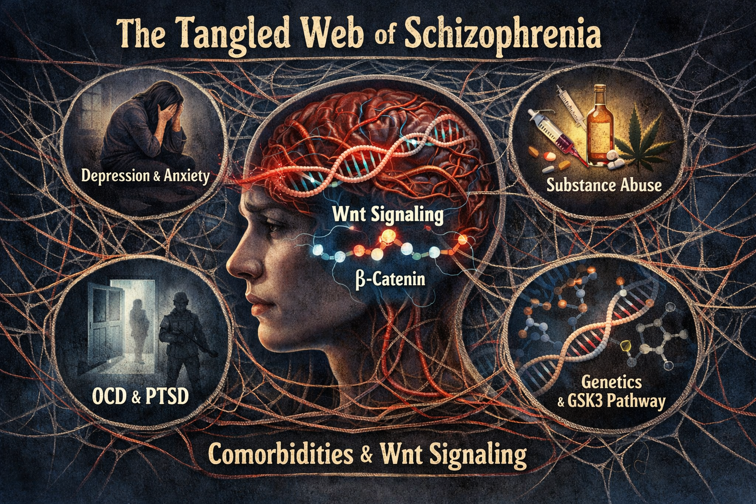
Cover Image Artstract designed by Cayley Borrud with the use of ChatGPT

Adolescence is often a rocky time full of firsts and figuring out identity. But what if in that volatile time, a teen decides to try vaping? It’s wrapped in colorful packaging, advertised to youth and the most used Tabacco product in youths [4]. How bad could just trying it be?

Image sourced from Alamy

The Neuroscience of Addiction

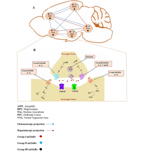
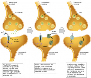
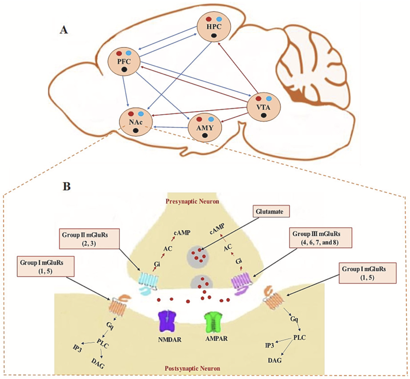
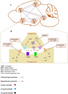
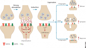
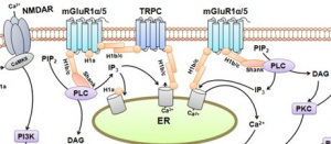
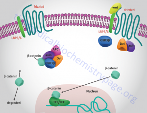
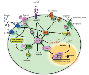
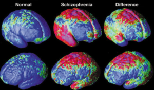
 E-cigarettes are designed to make the person crave more. It alters the brains reward center through the disruption of metabotropic glutamate receptors (mGluRs). The paper by Mozafari et. al explains how mGluRs are very involved in addiction. The absence of this vital receptor leads to increased glutamate. Glutamate is a neurotransmitter that drives the drug seeking behavior. It strengthens the circuits in the brain which would normally be a good thing however, the circuits that it is strengthening in this case would be the memories of the substance [1].

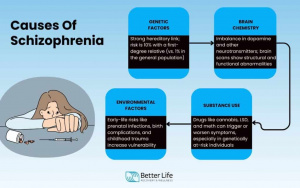
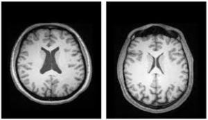
The brain could have structural changes such as increased dendritic spine density in the prefrontal cortex that can contribute to the formation of the ‘memory’ of vaping [3]. This is particularly a problem because it can make adolescents more likely to develop more addictions due to that rewiring of the brain. It also makes it much harder to stop their addiction since a non-fully developed brain is more likely to develop permanent addictions that follow them well into their adulthood [5].

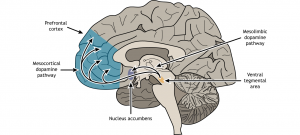
Image sourced from Baker Institute

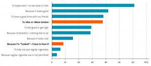
What started as curiosity driven experimentation would now be an addiction. This addiction can happen faster in teens than adults and it can greatly affect attention, impulse control and anxiety. The brain chemistry of the developing brain can change so rapidly that before nicotine is even consumed daily, withdrawal symptoms can appear [4].

The brain circuits involved in the development of addictions are constantly changing during adolescence which can lead to more risky behaviors [5]. What started as experimenting with e-cigarettes can quickly turn into experimentation and abuse of drugs and alcohol.

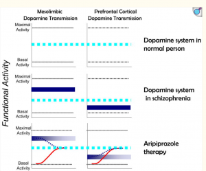
In adolescence, the dopamine signaling pathway is particularly sensitive and this pathway doesn’t reach full maturity until after adolescence [2]. This means that when a substance such as nicotine is taken, there would be a stronger reinforcement and quicker learned addictive behaviors [5]. The teenage brain is creating associations that vaping means reward and that association is the memory that’s hard to forget.

Now, let’s say, that teenager was able to stop vaping with the help of therapy and family support. That addictive pathway is still there and ready to be activated in moments of vulnerability or even from environmental cues associated with that nicotine memory [4]. Their brain is permanently changed. Unfortunately, adolescents don’t generally know that their actions are permantely changing their brain.

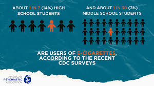
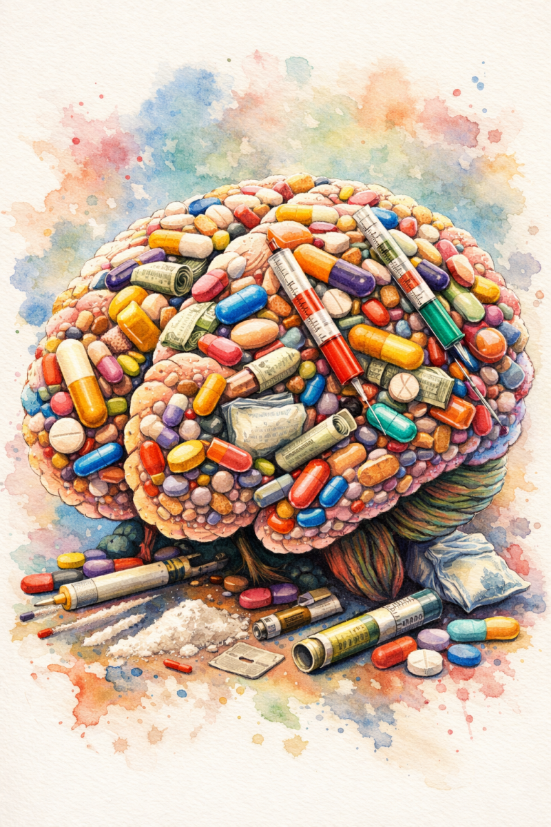
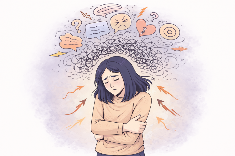
Image sourced from The American Psychiatric Association

Teens are biologically primed to become addicted therefore there needs to be more awareness and actions taken to protect them. There needs to be an urgency to protect teens before experimentation turns into long term consequences. By prioritizing prevention, regulation, and education, we can help protect developing brains during one of the most vulnerable stages of life.

Sources:

  1. Mozafari, R., Karimi-Haghighi, S., Fattahi, M., Kalivas, P., & Haghparast, A. (2023). A review on the role of metabotropic glutamate receptors in neuroplasticity following psychostimulant use disorder.Progress in Neuro-Psychopharmacology & Biological Psychiatry124, 110735. https://doi.org/10.1016/j.pnpbp.2023.110735
  2. Suri, D., Zanni, G., Mahadevia, D., Chuhma, N., Saha, R., Spivack, S., Pini, N., Stevens, G. S., Ziolkowski-Blake, A., Simpson, E. H., Balsam, P., Rayport, S., & Ansorge, M. S. (2023). Dopamine transporter blockade during adolescence increases adult dopamine function, impulsivity, and aggression.Molecular Psychiatry28(8), 3512–3523. https://doi.org/10.1038/s41380-023-02194-w
  3. Brown, R. W., & Kolb, B. (2001). Nicotine sensitization increases dendritic length and spine density in the nucleus accumbens and cingulate cortex.Brain Research899(1–2), 94–100. https://doi.org/10.1016/s0006-8993(01)02201-6
  4. (2024, October 17).E-cigarette use among youth. Smoking and Tobacco Use. https://www.cdc.gov/tobacco/e-cigarettes/youth.html
  5. Adolescents are neurologically more vulnerable to addictions | yale news. (2003, June 18). https://news.yale.edu/2003/06/18/adolescents-are-neurologically-more-vulnerable-addictions


Rewired Learning: The Hidden Science of Addiction

Featured image created by Julia Wolf and Microsoft CoPilot

The Science of Addiction

Addiction is no longer understood as simply a behavioral problem, it is a disorder rooted in long-lasting changes in the brain’s rewiring. Psychostimulant drugs like cocaine, amphetamine, and nicotine target the brain’s reward system, a network that includes the ventral tegmental area (VTA), nucleus accumbens (NAc), prefrontal cortex (PFC), amygdala, and hippocampus. These regions normally work together to guide behavior towards rewarding experiences like food, social interaction, and survival. Repeated drug exposure hijacks this system, strengthening pathways associated with the drug and making drug-seeking behavior automatic and persistent.

Figure 1. Key brain regions involved in reward and learning (top) communicate through glutamate pathways, while different types of glutamate receptors, including metabotropic glutamate receptors (mGluRs), regulate synaptic activity at the cellular level (bottom) [1].
At the heart of addiction is glutamate, the brain’s main excitatory neurotransmitter. Glutamate drives learning, memory, and synaptic plasticity. Synaptic plasticity is the ability of neurons to strengthen or weaken their connections based on experience. Psychostimulants disrupt normal glutamate signaling, elevating extracellular glutamate and altering receptor functioning. Over time, this rewiring leads to structural and functional changes, including more dendritic spines and stronger synaptic connections in key reward circuits. These changes make drug-related cues extremely powerful, driving craving and relapse even after long periods of abstinence.

A major player in these changes is the family of metabotropic glutamate receptors (mGluRs). These G-protein coupled receptors regulate longer-term, slower changes in synaptic strength. In particular, Group 1 mGluRs (mGluR1 and mGluR5) influence extracellular calcium levels, enzyme activity, and downstream signaling pathways, shaping learning and memory processes. Activation of these receptors can reinforce drug-seeking behavior, while blocking them can reduce craving and relapse. These interactions show that mGluRs have a central role in addiction-related neuroplasticity.

To learn more about the science of addiction, click here.

Long-Term Depression (LTD): The Brain’s Way of Letting Go

Long-term depression, or LTD, is the brain’s way of intentionally weakening synaptic connections. While long-term potentiation (LTP) strengthens certain pathways, LTD removes or diminishes connections that are no longer useful, helping the brain maintain balance and adapt to new information. LTD is essential for fine-tuning neural networks, pruning irrelevant memories, and preventing overexcitation that could damage neurons [2].

LTD occurs at excitatory synapses in key brain areas such as the hippocampus, cortex, striatum, and cerebellum. In the hippocampus, LTD helps clear out old or irrelevant memories, ensuring that new learning isn’t cluttered by outdated information. In the cerebellum, it fine-tunes motor coordination, helping us master smooth, precise movements. It also happens in other regions like the neocortex, striatum, and amygdala, where it contributes to emotion regulation, decision-making, and habit formation [4].

Figure 2. Neurons strengthen or weaken their connections over time: repeated activity adds more receptors and boosts signaling (LTP), while less activity removes receptors and reduces communication (LTD) [3].

There are different forms of LTD. Homosynaptic LTD weakens the synapse that is directly activated, while heterosynaptic LTD can weaken neighboring synapses that weren’t directly stimulated, helping the brain maintain balance across its networks [5]. At the molecular level, most LTD mechanisms involve removing AMPA receptors from the postsynaptic membrane, which reduces the strength of the synapse. In NMDA receptor-dependent LTD, common in the hippocampus, low-frequency signals from presynaptic neurons allow a small influx of calcium into the postsynaptic neuron, which activates enzymes that pull AMPA receptors out of the membrane. This results in a long-term decrease in synaptic strength, helping the brain fine-tune connections after individual events [6].

Another form, mGluR-dependent LTD, is found in both the hippocampus and cerebellum. Here, metabotropic glutamate receptors detect glutamate and trigger a cascade of signals inside the neuron, eventually removing AMPA receptors. This process is slower than NMDA-LTD but is important for adapting to prolonged experiences, such as repeated learning tasks or chronic drug exposure [7].

LTD isn’t just about pruning; it is very connected to memory, learning, and addiction. By eliminating unnecessary synapses, LTD supports efficient memory storage and refines motor skills. Disruption of mGluR-dependent LTD in reward pathways can make drug-related cues overly strong, contributing to addiction. Studying LTD helps us understand how the brain balances strengthening important connections while removing the ones we don’t need, offering insights into memory, motor learning, and potential therapies for addiction and neurological disorders [4].

To learn more about LTD functions and mechanisms, watch this short video!

Why This Matters: Addiction as a Failure to Adapt

Addiction can be seen as a form of maladaptive learning, a problem that is increasingly relevant in today’s society, not just with drugs, but also with behaviors like excessive phone use, social media, and gaming. The brain’s natural ability to adapt, strengthening useful pathways while weakening those that aren’t needed, can be redirected in harmful ways. Circuits linked to addictive behaviors become stronger and more dominant, while the brain struggles to weaken or “forget” behaviors that are harmful.

Studying mechanisms like long-term depression (LTD) and the role of mGluRs helps explain why cravings are so persistent and why relapse is common. This scientific understanding also highlights potential paths for treatment: therapies that restore the brain’s ability to weaken harmful connections or rebalance glutamate signaling may help people regain control over their behavior. Addiction isn’t just about desire; it’s about the brain being caught in a pattern it can’t easily reset and understanding this can guide more effective interventions.

References

[1] R. Mozafari, S. Karimi-Haghighi, M. Fattahi, P. Kalivas, and A. Haghparast, “A review on the role of metabotropic glutamate receptors in neuroplasticity following psychostimulant use disorder,” Progress in Neuro-Psychopharmacology and Biological Psychiatry, vol. 124, p. 110735, Jun. 2023, doi: https://doi.org/10.1016/j.pnpbp.2023.110735.

[2] D. Purves et al., “Long-Term Synaptic Depression,” Neuroscience. 2nd edition, 2001, Available: https://www.ncbi.nlm.nih.gov/books/NBK10899/

[3] Lumen Learning, “Synaptic Plasticity | Biology for Majors II,” Lumenlearning.com, 2008. https://courses.lumenlearning.com/wm-biology2/chapter/synaptic-plasticity/

[4] C. Lüscher and K. M. Huber, “Group 1 mGluR-Dependent Synaptic Long-Term Depression: Mechanisms and Implications for Circuitry and Disease,” Neuron, vol. 65, no. 4, pp. 445–459, Feb. 2010, doi: https://doi.org/10.1016/j.neuron.2010.01.016.

[5] Z. I. Mannan, S. Azam, R. K. Budhathoki, M. Nur Alam, and H. Kim, “Modeling homosynaptic and heterosynaptic plasticity with a single neuromemristive synapse,” Journal of Advanced Research, Oct. 2025, doi: https://doi.org/10.1016/j.jare.2025.10.031.

[6] D. Saal and R. C. Malenka, “The role of synaptic plasticity in addiction,” Clinical Neuroscience Research, vol. 5, no. 2–4, pp. 141–146, Nov. 2005, doi: https://doi.org/10.1016/j.cnr.2005.08.009.

[7] S. J. Kang and B.-K. Kaang, “Metabotropic glutamate receptor dependent long-term depression in the cortex,” The Korean Journal of Physiology & Pharmacology, vol. 20, no. 6, p. 557, 2016, doi: https://doi.org/10.4196/kjpp.2016.20.6.557.

When Abstaining Isn’t Enough: Psychostimulants and the Brain


Artstract 1 — Gannon Kuehn with assistance from AI


Out of the various brain-related afflictions discussed in these posts, addiction is unique in that it doesn’t arise from faulty wiring, but rather from something external. This is primarily why it might not be as charitably framed in our society. Addiction is more often seen as a moral failing, a choice — something that willpower alone should fix. Those framings have real consequences: they shape how families respond to loved ones in crisis, how courts sentence people who relapse, and how little urgency surrounds the search for effective treatments. For psychostimulants — cocaine, amphetamine, methamphetamine, nicotine — there are still no FDA-approved medications.

The scientific picture tells a different story. Addiction is a chronic, relapsing disorder driven by lasting changes in brain circuitry: changes that affect memory, motivation, impulse control, and the receptors that regulate how neurons communicate. Most of those changes don’t reverse when someone stops using. Some get worse. The assumption that the longer someone stays clean, the safer they are from relapse turns out to be wrong in important ways.

A 2023 review published in Progress in Neuropsychopharmacology & Biological Psychiatry examined how a family of receptors called metabotropic glutamate receptors (mGluRs) reshape the reward circuit during and after psychostimulant use. [1] What emerges is a picture of a brain that doesn’t simply reset when drug use stops; instead, it keeps changing, often in ways that increase vulnerability to relapse.

The receptor system nobody talks about

Most public conversation about addiction focuses on dopamine, but glutamate (the brain’s primary excitatory neurotransmitter) is equally central to why addiction is so hard to escape. Glutamatergic circuits connect the prefrontal cortex, the nucleus accumbens (NAc), the amygdala, and the ventral tegmental area: the core of the brain’s reward system. The mGluRs that sit throughout this circuit act as volume knobs, tuning the strength of signals flowing between these regions.

Figure 1: the location of Group I, II, and III receptors on both the pre- and postsynaptic neuron.

The review identifies three groups of mGluRs, each playing a distinct role. Group I receptors (mGluR1 and mGluR5), found mostly on the postsynaptic side, drive forms of synaptic strengthening and weakening that underlie learning and memory. Group II and III receptors sit primarily on presynaptic terminals and act as brakes, reducing glutamate release (Figure 1). [1] Psychostimulants like the drugs described in the first paragraph disrupt all of them.

What abstinence looks like

Consider someone who has been clean for two months. By most accounts, they are doing everything right and are on the road to recovery. The biology suggests, however, that they may actually be at greater risk of relapse than they were the week they quit. This phenomenon, the reason why the risk of relapse increases with time, is called the incubation of craving and was first documented in 1986. [2]  Since then, it has been confirmed across cocaine, methamphetamine, heroin, alcohol, and nicotine. 

The mGluR research helps explain why. During extended withdrawal, calcium-permeable AMPA receptors accumulate in NAc synapses, hardwiring drug-associated memories deeper into the circuit. [3] This is partly driven by a decline in mGluR1 activity. When researchers restore mGluR1 function, that accumulation reverses and incubated craving decreases, suggesting mGluR1 normally acts as a brake that gradually fails during abstinence. [4] At the same time, the prefrontal cortex remains persistently dysregulated in imaging studies of people in protracted withdrawal, undermining the impulse control needed to resist when a cue appears.

What this means

The review is careful to frame mGluRs as potential therapeutic targets precisely because the current situation is untenable: there are no FDA-approved medications for psychostimulant use disorder. The mGluR system offers multiple intervention points, from dampening cue reactivity to restoring glutamate homeostasis to interrupting the synaptic changes that incubate craving. But the broader implication is a clinical and cultural one. Telling someone that time and willpower are sufficient is not just incomplete — it runs counter to the neuroscience. The brain changes that drive relapse are not signs of weakness or poor motivation; they are the predictable outcome of a system that was, at the molecular level, systematically rewritten. Recovery is not as simple as merely abstaining, and understanding why is the first step toward correction.


(1) Mozafari, R.; Karimi-Haghighi, S.; Fattahi, M.; Kalivas, P.; Haghparast, A. A Review on the Role of Metabotropic Glutamate Receptors in Neuroplasticity Following Psychostimulant Use Disorder. Prog. Neuropsychopharmacol. Biol. Psychiatry 2023, 124, 110735. https://doi.org/10.1016/j.pnpbp.2023.110735. 

(2) Gawin, F.; Kleber, H. Pharmacologic Treatments of Cocaine Abuse. Psychiatr. Clin. North Am. 1986, 9 (3), 573–583. 

(3) Conrad, K. L.; Tseng, K. Y.; Uejima, J. L.; Reimers, J. M.; Heng, L.-J.; Shaham, Y.; Marinelli, M.; Wolf, M. E. Formation of Accumbens GluR2-Lacking AMPA Receptors Mediates Incubation of Cocaine Craving. Nature 2008, 454 (7200), 118–121. https://doi.org/10.1038/nature06995. 

(4) Loweth, J. A.; Scheyer, A. F.; Milovanovic, M.; LaCrosse, A. L.; Flores-Barrera, E.; Werner, C. T.; Li, X.; Ford, K. A.; Le, T.; Olive, M. F.; Szumlinski, K. K.; Tseng, K. Y.; Wolf, M. E. Synaptic Depression via mGluR1 Positive Allosteric Modulation Suppresses Cue-Induced Cocaine Craving. Nat. Neurosci. 2014, 17 (1), 73–80. https://doi.org/10.1038/nn.3590

The difficulty of unlearning: Addiction as a memory

Artstract by A. Griffith – Created with OpenAi

In the United States, 58.3% of the population used tobacco products, vaped nicotine, drank alcohol, or used an illicit drug within the previous month of the Substance Abuse and Mental Health Services Administration survey. [1] Addiction is prevalent and an important topic of treatment research, especially considering the physical and mental health impairments that accompany it. There are various theories of addiction and what is happening in the brain, the etiology, what causes behaviors, etc. When thinking about addiction, we often focus on reward. Dopamine and reward are a large part of what is occurring, but at its core addiction is an issue with learning and memory. Mechanistically in the brain, glutamate is largely involved in learning and memory implicating it in addiction as well. [2]

Addiction & Substance Use Disorder

Substance Use Disorder (SUD) is characterized by difficulty to limit drug intake and chronic relapsing. Psychostimulant Use Disorder (PUD) is a SUD with specifically psychostimulants, which include, cocaine, nicotine, and amphetamine-like drugs (methamphetamine). Drugs increase the dopamine present in the nucleus accumbens which results in tolerance, sensitization, and adaptation. There are no FDA-confirmed medications for the treatment of these disorders. [2]

Dopamine and Reward

The reward circuitry in the brain motivates drug use. Drug abuse physically alters brain connectivity, receptor and transporter expression of dopamine, and the synthesis and release of neurotransmitters involved in the reward circuit, including dopamine. Psychostimulants affect dopamine in multiple ways: binding to dopamine receptors, increasing dopamine release, inhibiting reuptake of dopamine, and decreasing the metabolism of dopamine. All of these cause more dopamine to be present and contribute to the “high” feeling that is sought in the reward cycle, as well as more of the substance being needed to have the same effect as it is continued to be used. [2]

Fig 1. Dopamine is released from the ventral tegmental area and the substantial nigra, then travels to the nucleus accumbens and prefrontal cortex. [3]

Glutamate

Glutamate is essential for synaptic plasticity, or the ability of the brain to form new connections, which is a large part of learning and memory. Receptors of the neurotransmitter glutamate include the inotropic receptors NMDA, AMPA, and kainite and the metabotropic receptors, or mGluRs, which are divided into three groups (Group I, Group II, Group III). [2]

Fig 2. Metabotropic Glutamate Receptors in the reward system [2]
Group I

Group I mGluRs are primarily located on postsynaptic neurons. They interact with the Gq protein in G protein-coupled receptors and lead to the second messengers IP3 and DAG. In some cases, blocking these receptors decreases drug seeking behaviors. [2]

Group II & III

Group II and III mGluRs are primarily located on presynaptic neurons and lead to the inhibition of adenylyl cyclase, and therefore the second messenger cAMP. They modulate neurotransmission, decreasing the chances of neurotransmitter release. This means they decrease the chance of dopamine being released, so agonists of these receptors have been implicated in reducing relapse. [2]

As part of the learning process, drug abuse affects the synaptic strength of neurons. This includes how neurons are physically wired together, as well as the receptors they express in the membrane; synaptic strength is modulated by glutamate. In drug abuse, the brain actually strengthens synapses to prepare to receive the same signal again. This is memory formation – the brain is actually rewired to “want” the drug again and become dependent on it. This increased synaptic strength is connected to relapse. [2]

Studies have shown that inhibiting glutamatergic inputs, blocking specific group I mGluRs, and agonists of group II & potentially group III mGluRs in animal models have prevented relapse and decreased drug seeking behaviors. The connection to glutamate and learning makes modulating glutamatergic neurotransmission a promising treatment for SUDs, particularly for the associated cognitive symptoms. Addiction is the brain learning from drug intake and forming memories to prepare for it to happen again, therefore, targeting this learning system is a potential to break the cycle of addiction and relapse.

Footnotes

[1] SAMHSA Releases Annual National Survey on Drug Use and Health. (2025, July 28). https://www.samhsa.gov/newsroom/press-announcements/20250728/samhsa-releases-annual-national-survey-on-drug-use-and-health

[2] Mozafari, R., Karimi-Haghighi, S., Fattahi, M., Kalivas, P., & Haghparast, A. (2023). A review on the role of metabotropic glutamate receptors in neuroplasticity following psychostimulant use disorder. Progress in Neuro-Psychopharmacology and Biological Psychiatry, 124, 110735. https://doi.org/10.1016/j.pnpbp.2023.110735

[3] Henley, C. (2021). Motivation and Reward. https://openbooks.lib.msu.edu/neuroscience/chapter/motivation-and-reward/


When Learning Goes Wrong: The Neuroscience of Addiction

Feature Image: Artstract by Emma Doty using AI

Overview: 

Psychostimulant use disorder (PUD) is a chronic, relapsing condition characterized by compulsive drug use despite harmful consequences. The review article (access here) emphasizes that addiction is not simply a behavioral issue, but a disorder rooted in long-lasting changes in brain circuitry. Specifically, the paper highlights how psychostimulants such as cocaine, amphetamine, and nicotine alter glutamatergic signaling within the brain’s reward system.[1]

Role of Glutamate:

At the center of this process is glutamate, the brain’s primary excitatory neurotransmitter. The article explains that psychostimulant use disrupts normal glutamate transmission and receptor function, particularly involving the metabotropic glutamate receptors (mGluRs). These disruptions lead to neuroadaptations that alter synaptic plasticity and reinforce drug-seeking behaviors. These changes remain even without the presence of the drug which contributes to relapses.[2]

Learning and Memory Impact:  

Importantly, the paper frames addiction as a form of maladaptive learning, as glutamate is essential in learning and memory processes. Drug-related experiences become ingrained in the brain’s reward circuitry, including regions like the nucleus accumbens, prefrontal cortex, and amygdala. Strong memories are formed that associate the stimulant with a feeling of pleasure which rewires the brain to seek these drugs. Over time, these changes alter how individuals respond to rewards, stress, and environmental cues, making it increasingly difficult to break the cycle of addiction.[3]

Long-Term Potentiation:  

To fully understand how these long-lasting changes occur, it is essential to consider long-term potentiation (LTP). LTP refers to the persistent strengthening of synapses following a pattern of intense activity and is widely regarded as the cellular basis of learning and memory.[4]

This intense neuronal activity leads to the release of large amounts of glutamate, which signifies the beginning of this process. Glutamate then activates AMPA receptors, causing depolarization of the postsynaptic neuron. When this depolarization is strong enough, it removes the magnesium block from NMDA receptors, allowing a rapid influx of calcium. This acts as a molecular trigger, activating proteins such as CaMKII, which promote the insertion of additional AMPA receptors into the postsynaptic membrane. These extra receptors are inherently more sensitive to glutamate. As a result, the synapse becomes more responsive to future signals, strengthening the connection between neurons.[5]

Image of LTP Mechanism: https://www.researchgate.net/figure/nduction-and-expression-of-LTPStrong-repetitive-stimulation-of-the-input-to-a_fig2_12895506

LTP occurs in two phases: an early phase (E-LTP), which occurs independently of protein synthesis, and a late phase (L-LTP), which requires gene expression and structural changes to the synapse. During this late phase, transcription factors like CREB drive the growth of dendritic spines and the expansion of synaptic contact area, allowing these strengthened connections to persist for hours, days, or even longer.[6]

Final Thoughts:

In the context of addiction, LTP provides an explanation for the role of learning and memory. Psychostimulants effectively hijack the brain’s natural learning processes by inducing LTP within reward circuits. Each drug exposure strengthens the neural pathways associated with drug use, making those pathways more easily activated in the future.

Ultimately, the article emphasizes that targeting glutamatergic signaling and synaptic plasticity, such as the LTP mechanism, may be key to developing effective treatments for addiction. By disrupting or reshaping these maladaptive neural connections, it may be possible to weaken the hold that drug-related memories have on behavior.

[1] Mozafari et al., “A Review on the Role of Metabotropic Glutamate Receptors in Neuroplasticity Following Psychostimulant Use Disorder,” Progress in Neuro-Psychopharmacology and Biological Psychiatry 124 (June 2023): 110735, https://doi.org/10.1016/j.pnpbp.2023.110735.

[2] Mozafari et al., “A Review on the Role of Metabotropic Glutamate Receptors in Neuroplasticity Following Psychostimulant Use Disorder.”

[3] Mozafari et al., “A Review on the Role of Metabotropic Glutamate Receptors in Neuroplasticity Following Psychostimulant Use Disorder.”

[4] “What Is Long-Term Potentiation and How Does It Work?,” ScienceInsights, November 21, 2025, https://scienceinsights.org/what-is-long-term-potentiation-and-how-does-it-work/.

[5] ScienceInsights, “What Is Long-Term Potentiation and How Does It Work?”

[6] “Long-Term Potentiation – an Overview | ScienceDirect Topics,” accessed March 24, 2026, https://www.sciencedirect.com/topics/neuroscience/long-term-potentiation.

Addiction as Memory: When Psychostimulants Cause Learning to Become a Problem

Image from Clay Behavior Health Center (https://ccbhc.org/understanding-addiction/)

Psychostimulants Spark Learning Pathways

Your brain learns from experience, remembers what feels good, and craves those behaviors.  Every time you eat, socialize, or achieve something, your brain strengthens connections to help you do it again. This learning pathway relies on glutamate, the brain’s main excitatory neurotransmitter, responsible for most communication between neurons. However, the same system that helps you learn healthy behaviors is also influenced by various substances. Therefore, when something overstimulates this system, such as drug use, the brain remembers the feeling and learns it. Psychostimulants, including cocaine, nicotine, and methamphetamine, flood the brain with dopamine [1]. This creates intense feelings of reward and alters glutamate signaling, locking that experience into memory.

How Glutamate Leads to Learning & Memory

Glutamate receptors are essential for how the brain learns and forms memories because they control how strongly neurons communicate and form synapses. NMDA receptors initiate long-term changes in the brain by allowing calcium into cells, triggering processes needed for learning. AMPA receptors strengthen these connections by increasing fast signaling between neurons, making memories more stable. Kainate receptors help regulate overall brain activity, supporting this communication. Metabotropic receptors (mGluRs) play more specific roles in this system, and are divided into 3 main groups. Group I mGluRs enhance signaling and modulate excitatory signaling, strengthening learning pathways. Group II mGluRs are important for the reward system, and regulate neurotransmitter release. Group  III mGluRs act as brakes by suppressing glutamate release and preventing overstimulation [1]. Together these receptors balance strengthening and controlling brain signals, allowing normal learning and memory to occur. But when this normal balance is disrupted by psychostimulant use, it leads to overly strong and persistent memories like those seen in addiction.

Receptor What It Normally Does What Happens in Addiction
NMDA Helps with learning & memory Strengthens drug-related memories
AMPA Sends fast brain signals Boosts cravings & drug sensitivity
Kainate Helps control brain activity Supports excitation in addiction
Group I (mGluR1, 5) Boosts brain signaling and learning Increases drug-seeking & relapse
Group II (mGluR2, 3) Reduces glutamate release Decreases cravings & relapse
Group III (mGluR4,6,7,8) Slows down brain signaling Control addiction signals
Table 1: This table shows how different glutamate receptors effect addiction, either by strengthening behaviors or helping regulate and reducing drug-seeking behavior [1].

Helpful Organizers: Homer Proteins

Within your brain, there are scaffolding proteins called Homer proteins that help organize communication between receptors [1]. In the glutamate pathway, Homer proteins regulate group I mGluR signaling to help with excitatory neurotransmission and synaptic plasticity [2], the process in the brain that results in learning and memory. They help with structure and keeping signaling systems stable and efficient. By controlling glutamate signaling, Homer proteins indirectly influence dopamine reward pathways as well [4]. Therefore, they are important in the signaling that effects learning and feelings of reward.

Figure 1: Long-form Homer proteins (peach rectangles, “H1b/c”) stabilize mGluR signaling by linking receptors to intracellular calcium pathways, while short-form Homer proteins (peach circles, “H1a”) disrupt this scaffolding, altering signaling and promoting synaptic changes [5].

Rewiring the Brain & Relapse

Synaptic plasticity involves connections between neurons strengthen or weaken over time, altering how we learn and form memories. Processes like long-term potentiation (LTP) and long-term depression (LTD) shape how memories are formed. Drugs disrupt the balance in this process. Instead of normal learning, they exaggerate certain pathways, particularly those tied to reward. Chronic psychostimulant use increases a type of homer protein, Homer1a, which disrupts the normal scaffolding with mGluR receptors and makes glutamate signaling unstable [3].

Therefore, use of psychostimulants causes glutamate levels and synaptic connections to change, allowing the brain to undergo drug-induced neuroplasticity. This neuroplasticity causes the memory system to become sensitive to cues related to drug use. Places, feelings, or another memory can trigger intense craving. Drug-seeking is driven by strengthened neural pathways that have been biologically reinforced. Glutamate signaling can increase when exposed to triggers, reactivating the learned pathways and can result in relapse [6].

Conclusion

Addiction involves deep changes in how the brain learns, remembers, and prioritizes behavior. Understanding glutamate signaling and Homer proteins helps identify the root cause and gives researchers new targets for treatment. Therefore, future therapies may focus on helping the brain unlearn harmful patterns. The brain’s adaptability, learning, and memory systems are what allows addiction to be such a strong habit.

  1. Mozafari, R., Karimi-Haghighi, S., Fattahi, M., Kalivas, P., Haghparast, A. 2022. A review on the role of metabotropic glutamate receptors in neuroplasticity following psychostimulant use disorder. Frontiers in Pharmacology, 124. https://doi.org/10.1016/j.pnpbp.2023.110735
  2. Shiraishi-Yamaguchi, Y., Furuichi, T. The Homer family proteins. Genome Biol 8, 206 2007. https://doi.org/10.1186/gb-2007-8-2-206
  3. Niswender, C. M., & Conn, P. J. 2010. Metabotropic glutamate receptors. Annual Review of Pharmacology and Toxicology. 10.1146/annurev.pharmtox.011008.145533
  4. Szumlinski, K. K., Ary, A. W., & Lominac, K. D. 2008. Homers regulate drug-induced neuroplasticity: implications for addiction. Biochemical pharmacology, 75(1), 112–133. https://doi.org/10.1016/j.bcp.2007.07.031
  5. Peng Luo, Xia Li, Zhou Fei, Waisang Poon. 2012. Scaffold protein Homer 1: Implications for neurological diseases, Neurochemistry International. Volume 61, Issue 5, Pages 731-738. https://doi.org/10.1016/j.neuint.2012.06.014.
  6. Ménard, C., & Quirion, R. 2012. Group 1 Metabotropic Glutamate Receptor Function and Its Regulation of Learning and Memory in the Aging Brain. Frontiers in Pharmacology, 3. https://pubmed.ncbi.nlm.nih.gov/23091460/

Antipsychotics: A Balance of Safety and Comfort

Schizophrenia is one of the hardest mental conditions to treat and it can often be difficult to get patients to comply with a medication regiment. This is due to a relatively simple reason, that reason being that antipsychotics can make those who take it feel absolutely miserable. This is something that is well known by pharmaceutical companies and this has resulted in different generations of antipsychotics that each have their pros and cons. But to understand how these medications work we must understand how schizophrenia works.

How Schizophrenia Works

Schizophrenia has long been considered an issue with dopamine (DA), and this can be explained by the symptoms. The symptoms of schizophrenia have been associated with D2 receptor misfiring in the mesolimbic pathway leading to hallucinations, delusions and disorganized thinking. This has been supported through studies that show increasing DA release can induce psychosis and blocking DA relieves it. More recently, the hypothesis has changed from too much DA to DA dysregulation, with too much DA in some areas and not enough in others (1). This change in philosophy can also be seen with the mechanisms of third generation antipsychotics, which will be explained further on in this post. For more details on the DA hypothesis, check out this article.

Researchers have recently suggested schizophrenia is resulted from dysregulated Wnt signaling caused by DA misfiring. The way Wnt signaling works is relatively simple, and one must start at the destruction complex to understand it. When Wnt signaling is not active, there is a destruction complex that leads to the destruction of B-catenin. B-catenin is a transcription factor that is associated with cell/neuron growth. One of the most important parts of this complex is called GSK3B, this is basically what marks B-catenin for destruction. When Wnt signaling is active, it leads to the disassociation of the destruction complex leading to B-catenin being able to enter the nucleus. The theory behind all this is too much DA signaling during development leads to Wnt dysregulation and the lack 0f B-catenin getting into the nucleus results in abnormal brain pathways associated with schizophrenia. Now you are probably wondering what DA’s role in this is, rather than DA itself it relates more to what DA receptor activation leads to. First we must talk about something called AKT, when this becomes activated it inhibits GSK3B therefore leading to more B-catenin accumulation. D2 receptor activation leads to the inhibition of AKT and therefore strengthens the destruction complex (2). This has been supported in animal models exploring the role of GSK3B and AKT in schizophrenia and how it relates to DA and it can also be supported when looking at the way some antipsychotics work. For more details on this hypothesis of schizophrenia check out this article.

This is a visual on Wnt signaling to help with understanding. With inactive signaling on the left, and active signaling on the right. Sourced from.

First Generation Antipsychotics (3)

First generation antipsychotics (FGA) were first offered in 1952. These can result in some very unpleasant symptoms such as sedation and extrapyramidal symptoms, this means symptoms similar to those of Parkinson’s such as movement issues like dyskinesia or tremors. There are two kinds of FGAs, high potency, which have lower sedative symptoms but higher extrapyramidal symptoms, and low potency, which result in higher sedation and lower extrapyramidal symptoms.

While the mechanisms of action vary between different drugs, the common factor is blocking of D2 receptors in the substantia nigra and the mesolimbic pathway. As discussed previously, reduced D2 signaling results in more Wnt signaling. Additionally, many FGAs directly interact with Wnt signaling. Clozapine induces phosphorylation of GSK-3B and AKT, both of these result in the disassociation of the destruction complex and therefore more B-catenin can accumulate. Lithium, another FGA, prevents inactivation of AKT and also directly inhibits GSK3B, which also leads to more B-catenin accumulation.

The side effects of FGAs are the most severe out of all of the antipsychotic generations. The most prevalent of these side effects are the extrapyramidal effects. These result from blocking of D2 receptors in the substantia nigra, an area very important for motor planning and execution. This area is affected by Parkinson’s, which is why we see similar symptoms between the two. Disruption of the mesolimbic pathway can result in social apathy and social withdrawal, turning the patient into a shell of themselves. Additionally, FGAs interact with histamine receptors, which induces sedation. These side effects are seen in some form in most people who take FGAs, and therefore further development of drugs with less side effects was needed.

Second Generation Antipsychotics (4)

Second generation antipsychotics (SGAs) were first offered in 1990 and have been found to result in less severe side effects. With SGAs D2 receptors are still blocked in the mesolimbic pathway, but not as much in the substantia nigra as in FGAs.  This results in less of the extrapyramidal symptoms seen in FGAs. Additionally, SGAs blocks serotonin receptors, primarily in the frontal cortex. This leads to more DA release which helps relieve some of the side effects caused by antipsychotics. SGAs have been found to increase Wnt signaling through D2 receptor blocking, similarly to FGAs. There is less evidence showing they are direct modulators similarly to FGAs like lithium, but through D2 receptors blocking, inherently increasing Wnt signaling.

Side effects of SGAs are similar to that of FGAs. The difference is seen in the decrease of extrapyramidal side effects. Due to less D2 blocking in the substantia nigra, less extrapyramidal side effects are seen in SGAs. There is still a risk of these side effects, but the risk is lower and the symptoms are also less severe than FGAs. SGAs also mess with histamine receptors, leading to sedation. Something unique to SGAs is they have been associated with weight gain and insulin resistance/type 2 diabetes. This is caused through serotonin receptor blockage, messing with the appetite and eating habits. While these side effects are a great improvement from FGAs, they are far from ideal. This has resulted in the development of more unique antipsychotics.

Third Generation Antipsychotics (5)

The mechanisms behind third generation antipsychotics (TGA) vary greatly, therefore, we will focus primarily on the most well studied one, aripiprazole. Aripiprazole was first offered in 2002 and does not work like other antipsychotics. Rather than blocking D2 receptors, it works by selectively firing them. What this means is that when there is too much DA, aripiprazole competes with it and binds to the receptor instead, acting as an antagonist in this context. When there is too little DA, aripiprazole picks up the slack, acting as an agonist in this context.

(Graph showing how aripiprazole acts depending on the context, source)

This functional selection of firing results in less side effects, and less severe side effects. The side effects are more or less the exact same just less risk of experiencing them and less severity. There are a few exceptions though. Something unique to TGAs is they actually can cause restlessness and insomnia rather than sedation due to continued firing of D2 receptors and less histamine receptor interaction.

The Future

As antipsychotics continue to be developed, schizophrenia treatment will continue to change and mold depending on the current literature. Antipsychotics have been around for 74 years, which in the grand scheme of things is very recently. There are people who developed schizophrenia before the discovery of antipsychotics still alive today, and while this fact can be depressing in a way, it can also be promising. They have come very far compared to how they started, and as treatment philosophy has started to shift from complete D2 receptor blocking to selective firing, the side effects will hopefully continue to improve.

(Source)


Feature image developed through Google Gemini 3 and Eli Hunt. Prompt “make an image that represents schizophrenia and the different generations of antipsychotics” followed by “make it more artistic, less words”

  1. Brisch, R., Saniotis, A., Wolf, R., Bielau, H., Bernstein, H. G., Steiner, J., Bogerts, B., Braun, K., Jankowski, Z., Kumaratilake, J., Henneberg, M., & Gos, T. (2014). The role of dopamine in schizophrenia from a neurobiological and evolutionary perspective: old fashioned, but still in vogue. Frontiers in psychiatry5, 47. https://doi.org/10.3389/fpsyt.2014.00047
  2. Singh K. K. (2013). An emerging role for Wnt and GSK3 signaling pathways in schizophrenia. Clinical genetics83(6), 511–517. https://doi.org/10.1111/cge.12111
  3. Grace, A. A., & Uliana, D. L. (2023). Insights into the Mechanism of Action of Antipsychotic Drugs Derived from Animal Models: Standard of Care versus Novel Targets. International journal of molecular sciences24(15), 12374. https://doi.org/10.3390/ijms241512374
  4. Chokhawala, K. P., & Stevens, L. (2023). Antipsychotic Medications. In StatPearls. StatPearls Publishing.
  5. Mailman, R. B., & Murthy, V. (2010). Third generation antipsychotic drugs: partial agonism or receptor functional selectivity?. Current pharmaceutical design, 16(5), 488–501. https://doi.org/10.2174/138161210790361461

The Silent Risk Factors of Schizophrenia

The Silent Risks of Schizophrenia

Schizophrenia is a mental health condition that is thought to be a disorder of brain development and neural connectivity [2]. Symptoms of Schizophrenia typically show up in one’s early twenties however, the origins of Schizophrenia can begin during brain formation as a fetus [1][2].

Maternal Immune Activation

One of the lesser-known risk factors of schizophrenia is Maternal Immune Activation (MIA). This occurs when a pregnant person experiences an infection and their immune system responds by releasing cytokines. Cytokines are protective signaling molecules that help with regulating immunity. However, these cytokines can cross through the placenta and affect brain development [3]. One cytokine in particular, interleukin-8, is linked to schizophrenia [4]. This exposure to cytokines can alter how neural circuits form, specifically the Wnt pathway, during critical periods of development [2].

It is commonly known that sickness causes immune response, but did you know that environmental factors such as pollution from highways and microplastics can also trigger an immune response? [5] This Immune activation occurs without noticeable illness which can pose a risk to the developing brain of the fetus.

Timing also matters. Immune activation poses more of a significant risk during the first and second trimester of pregnancy [8]. This is when foundational neural circuits and brain structures being formed. Even subtle disruptions during this time can have long-term effects.

Maternal immune response is also associated with other neurodevelopmental and neuropsychiatric conditions including ADHD and Autism spectrum disorder [6]. This suggests that there is a shared neurodevelopment pathway where similar disruptions can result in different outcomes depending on genetics and life experiences.

Image sourced from BetterLife

Genetic Vulnerability

Environmental factors do not act alone. Instead, they interact with one’s genetics to influence the risk of schizophrenia. Genetic variations involved in immune function, brain development and systems like Wnt/GSK3 signaling. While certain genetics can increase vulnerability, an environmental trigger would have to play a role for there to be alterations in brain development [7].

Genetics make up 80% of schizophrenia risk. A child with two affected parents has a 40% prevalence while a child with one affected parent has a 12% prevalence [9]. Changes in brain chemicals like glutamate and dopamine may also play a part in schizophrenia prevalence [10].

Image sourced from PatientsEngage

Changing How We Think About Schizophrenia

Schizophrenia is not a sudden or random condition. It occurs because of brain development and genetics before symptoms appear.

Many of these risk factors are out of one’s control since processes like immune activation are normal and necessary to protect the body. This shows that schizophrenia is not a result of personal failures. It’s important that as a society, we create more understanding to help reduce the stigma.

What appears later in life reflects subtle changes that had occurred in brain development [2]. By understanding these hidden risks, we can move closer to understanding how the brain develops to develop prevention measures and treatments.

Sources:

  1. Schizophrenia: Symptoms, causes & treatment options | nami. (n.d.). Retrieved March 25, 2026, from https://www.nami.org/types-of-conditions/schizophrenia/#:~:text=Although%20schizophrenia%20can%20occur%20at%20any%20age%2C,is%20possible%20to%20live%20well%20with%20schizophrenia
  2. Singh, K. K. (2013). An emerging role for Wnt and GSK3 signaling pathways in schizophrenia.Clinical Genetics83(6), 511–517. https://doi.org/10.1111/cge.12111
  3. Ayoub, G. (2025). Neurodevelopmental impact of maternal immune activation and autoimmune disorders, environmental toxicants and folate metabolism on autism spectrum disorder.Current Issues in Molecular Biology47(9), 721. https://doi.org/10.3390/cimb47090721
  4. Ellman, L. M., Deicken, R. F., Vinogradov, S., Kremen, W. S., Poole, J. H., Kern, D. M., Tsai, W. Y., Schaefer, C. A., & Brown, A. S. (2010). Structural brain alterations in schizophrenia following fetal exposure to the inflammatory cytokine interleukin-8.Schizophrenia Research121(1–3), 46–54. https://doi.org/10.1016/j.schres.2010.05.014
  5. Yang, W., Jannatun, N., Zeng, Y., Liu, T., Zhang, G., Chen, C., & Li, Y. (2022). Impacts of microplastics on immunity.Frontiers in Toxicology4, 956885. https://doi.org/10.3389/ftox.2022.956885
  6. Cattane, N., Richetto, J., & Cattaneo, A. (2020). Prenatal exposure to environmental insults and enhanced risk of developing Schizophrenia and Autism Spectrum Disorder: Focus on biological pathways and epigenetic mechanisms.Neuroscience & Biobehavioral Reviews, Prenatal Stress and Brain Disorders in Later Life117, 253–278. https://doi.org/10.1016/j.neubiorev.2018.07.001
  7. Wahbeh, M. H., & Avramopoulos, D. (2021). Gene-environment interactions in schizophrenia: A literature review.Genes12(12), 1850. https://doi.org/10.3390/genes12121850
  8. Mor, G., Cardenas, I., Abrahams, V., & Guller, S. (2011). Inflammation and pregnancy: The role of the immune system at the implantation site.Annals of the New York Academy of Sciences1221(1), 80–87. https://doi.org/10.1111/j.1749-6632.2010.05938.x
  9. Causes and risk factors of schizophrenia. (n.d.). Clínic Barcelona. Retrieved March 25, 2026, from https://www.clinicbarcelona.org/en/assistance/diseases/schizophrenia/causes-and-risk-factors
  10. Mayo Clinic. (n.d.). Schizophrenia: Symptoms and causes. Retrieved March 25, 2026, from https://www.mayoclinic.org/diseases-conditions/schizophrenia/symptoms-causes/syc-20354443

Cover image sourced from ChatGPT


The Tangled Web: Comorbidities, Wnt Signaling, and the Complexity of Schizophrenia


Comorbidities

Schizophrenia almost never arrives alone. In clinical practice, psychiatric comorbidities are usually the rule rather than the exception, and their prevalence is striking. Roughly 50% of patients experience significant depression at some point across the illness course, with PTSD and OCD corresponding to an estimated 29% and 23% prevalence among patients respectively. Furthermore, Substance use disorders carry a lifetime prevalence of approximately 47%, making them the most common comorbidity of all.1

That substance use figure deserves particular attention, as several competing explanations exist. Such explanations include mere chance, the use of substances as self-medication, substances directly precipitating psychosis, and shared neurobiological vulnerability. The self-medication hypothesis is intuitive but loses force in the era of better-tolerated second-generation antipsychotics as you would expect a decline in substance abuse rates – a decline which doesn’t exist. Interestingly, a meta-analysis suggested adolescent cannabis use as being linked to schizophrenia developing later in life.2 Nevertheless, any concrete link remains unsubstantiated. A few things cut across all of these comorbidities: each is associated with worse outcomes than schizophrenia alone, all appear across the full illness course, the neurobiological basis of each remains understudied, and treatment is largely trial and error without clear evidence-based guidance tailored to these subgroups.

Recent Work

Figure 1: The Canonical Wnt pathway turned off (left) and turned on (right)

This is where emerging molecular research becomes relevant not as a solution, but as a framework for asking better questions. A 2013 review by Karun Singh in Clinical Genetics synthesized converging evidence implicating Wnt and glycogen synthase kinase 3 (GSK3) signaling in the biological basis of schizophrenia.3 The canonical Wnt pathway regulates β-catenin stability and downstream gene transcription, and plays a prominent role in neural development. The pathway depends on the stability of β-catenin: whether it gets broken down or or survives long enough to enter the nucleus. GSK3β is the key enzyme that drives that breakdown, so when Wnt signaling is active, or when GSK3β is blocked, β-catenin accumulates and gene transcription follows. This is illustrated clearly in Figure 1, where Wnt signaling being turned off results in the destruction of β-catenin, and Wnt being turned on results in the surplus of β-catenin which enters the nucleus.4 When disrupted, the consequences affect the very systems implicated in psychiatric vulnerability.

The pharmacological evidence, as laid out by Singh, is striking. Lithium, one of psychiatry’s oldest empirically validated treatments, works partly by directly inhibiting GSK3β and stabilizing β-catenin, effectively activating canonical Wnt signaling.5 Additionally, antipsychotics, whose primary target is the dopamine D2 receptor, modulate GSK3β activity through the Akt pathway as a downstream effect. Even metabotropic glutamate receptor agonists, a non-dopaminergic avenue, regulate GSK3 and accumulate Wnt pathway proteins with repeated treatment. The fact that these three mechanistically distinct drug classes converge on the same signaling network is not coincidental.

Human genetic findings reinforce this further. When scientists look at the genes associated with schizophrenia, they keep finding the same pattern. A gene called DISC1, one of the most well-known schizophrenia risk genes, works by blocking the same protein that lithium blocks, suggesting that what lithium does as a drug, this gene does naturally, and when it goes wrong, the consequences look like schizophrenia.6 Studies examining brain tissue from people who had schizophrenia have also found reduced levels of a protein called Akt1, and large-scale genetic studies keep flagging the same region.7 Taken together, these findings suggest that disruptions in this particular signaling network aren’t just a side observation; instead, they may be a genuine part of what actually causes schizophrenia in the first place.

What does this mean for the comorbidity problem?

The high rates of depression, anxiety, OCD, and substance use disorders seen alongside schizophrenia may not be coincidental — they may reflect a shared underlying disruption in Wnt/GSK3 signaling that plays out differently across neural systems. Because this pathway touches so many aspects of brain development and function, a single disruption early on could leave multiple systems vulnerable in ways that manifest as distinct but co-occurring disorders. This doesn’t solve the comorbidity problem, but it suggests these conditions may share a common biological origin rather than simply co-occurring by chance.


(1) Buckley, P. F.; Miller, B. J.; Lehrer, D. S.; Castle, D. J. Psychiatric Comorbidities and Schizophrenia. Schizophr. Bull. 2009, 35 (2), 383–402. https://doi.org/10.1093/schbul/sbn135. 

(2) Moore, T. H.; Zammit, S.; Lingford-Hughes, A.; Barnes, T. R.; Jones, P. B.; Burke, M.; Lewis, G. Cannabis Use and Risk of Psychotic or Affective Mental Health Outcomes: A Systematic Review. The Lancet 2007, 370 (9584), 319–328. https://doi.org/10.1016/S0140-6736(07)61162-3. 

(3) Singh, K. K. An Emerging Role for Wnt and GSK3 Signaling Pathways in Schizophrenia. Clin. Genet. 2013, 83 (6), 511–517. https://doi.org/10.1111/cge.12111. 

(4) Xue, C.; Chu, Q.; Shi, Q.; Zeng, Y.; Lu, J.; Li, L. Wnt Signaling Pathways in Biology and Disease: Mechanisms and Therapeutic Advances. Signal Transduct. Target. Ther. 2025, 10 (1), 106. https://doi.org/10.1038/s41392-025-02142-w. 

(5) Klein, P. S.; Melton, D. A. A Molecular Mechanism for the Effect of Lithium on Development. Proc. Natl. Acad. Sci. U. S. A. 1996, 93 (16), 8455–8459. https://doi.org/10.1073/pnas.93.16.8455. 

(6) Mao, Y.; Ge, X.; Frank, C. L.; Madison, J. M.; Koehler, A. N.; Doud, M. K.; Tassa, C.; Berry, E. M.; Soda, T.; Singh, K. K.; Biechele, T.; Petryshen, T. L.; Moon, R. T.; Haggarty, S. J.; Tsai, L.-H. Disrupted in Schizophrenia 1 Regulates Neuronal Progenitor Proliferation via Modulation of GSK3beta/Beta-Catenin Signaling. Cell 2009, 136 (6), 1017–1031. https://doi.org/10.1016/j.cell.2008.12.044. 

(7) Emamian, E. S.; Hall, D.; Birnbaum, M. J.; Karayiorgou, M.; Gogos, J. A. Convergent Evidence for Impaired AKT1-GSK3beta Signaling in Schizophrenia. Nat. Genet. 2004, 36 (2), 131–137. https://doi.org/10.1038/ng1296.

The Making of the Mind: What Changes in Schizophrenia

The human brain is like a city under constant construction, with billions of cells building and connecting to form the networks that shape our thoughts, emotions, and perception of reality. This process depends on precise signals that tell cells where to go and what to do. Most of the time, everything comes together as it should, but when those signals are even slightly off, the brain can develop in ways that aren’t immediately visible yet have lasting effects on how the world is experienced. In conditions like schizophrenia, these early changes in brain development may play a key role in shaping how reality is perceived later in life.

The Science of Schizophrenia: Building (and Misbuilding) the Brain

At the center of brain development are powerful signaling systems that guide how neurons grow, connect, and communicate. One of the most important of these is the Wnt signaling pathway, which helps control how brain cells develop and organize into functional networks. A key part of this pathway involves a protein called β-catenin. When Wnt signaling is active, β-catenin builds up and turns on genes that support healthy brain development. When the pathway is less active, another protein called glycogen synthase kinase-3β (GSK-3β) helps in the breakdown of β-catenin, preventing those genes from being expressed. This balance between activation and breakdown is critical, if it is disrupted, brain development can be altered.

This pathway is also closely connected to dopamine, a neurotransmitter often linked to schizophrenia. Dopamine plays a major role in motivation, reward, and perception, and imbalances in dopamine signaling are strongly associated with symptoms such as hallucinations and delusions. Emerging research suggests that dopamine and Wnt signaling may interact, meaning that disruptions in one system can influence the other. For example, changes in dopamine activity can affect GSK-3, which in turn impacts the Wnt pathway and brain development. Together, these overlapping systems highlight how schizophrenia may arise not from a single issue, but from interconnected changes in both brain signaling and structure [1].

Figure 1. Diagram showing how the Wnt signaling pathway interacts with dopamine and other neurotransmitter systems in schizophrenia. Key regulators like GSK-3β are highlighted, along with how medications such as antipsychotics and lithium influence these pathways [1].
To learn more about Wnt signaling, dopamine, and their connection to schizophrenia, click here.

What Brain Imaging Reveals

While molecular pathways like Wnt help explain what may be happening at a cellular level, brain imaging techniques like fMRI, DTI, and PET scans show how these changes appear in the brain itself. These tools reveal that schizophrenia is not limited to a single area, but instead involves multiple interconnected regions that support thought, emotion, and perception [1].

One of the most consistently affected areas is the prefrontal cortex, which is responsible for planning, attention, working memory, and decision making. In individuals with schizophrenia, this region often shows reduced activity, known as hypofrontality, along with decreased gray matter and weaker connectivity. These changes are strongly linked to cognitive and negative symptoms, such as concentrating and organizing thoughts.

The amygdala and hippocampus, which are involved in emotion and memory, are also significantly affected. Abnormal amygdala activity can contribute to heightened emotional responses, paranoia, and difficulty interpreting social cues. The hippocampus, often smaller and functionally altered, plays a role in memory and distinguishing internal thoughts from external reality, helping explain symptoms like hallucinations and delusions in schizophrenia [1].

Disruptions are also seen in the thalamus, which normally acts as a relay center for sensory information. Reduced volume and impaired communication with cortical regions can lead to sensory misprocessing, hallucinations, and difficulty focusing attention. In addition, enlargement of the brain’s ventricles, one of the most consistent findings in schizophrenia, typically reflects a loss of surrounding brain tissue.

Figure 2. Comparison of brain affected by schizophrenia (left), showing enlarged ventricles, and a neurotypical brain (right) [4].
The striatum, particularly within the mesolimbic dopamine pathway, is involved in reward and motivation. Dysfunction in this region is associated with anhedonia, reduced motivation, and social withdrawal, which are negative symptoms of schizophrenia [1].

Beyond the regions discussed above, schizophrenia involves widespread changes throughout the brain. There is an overall decrease in both gray and white matter, leading to reduced connectivity between neurons. Decreased myelination slows communication between neurons, while increased brain inflammation may further disrupt signaling. Some research also suggests accelerated brain aging, with schizophrenic brains appearing 3.5 years older than neurotypical brains [3]. Together, these findings reflect that schizophrenia is a disorder of brain networks rather than a single isolated issue.

Figure 3. The image shows a neurotypical brain (right), a brain affected by schizophrenia (middle), and a comparison highlighting gray matter loss [5] .
If interested in learning more about brain regions and symptoms related to schizophrenia, click here for a video!

Why This Matters

Looking at schizophrenia through both a molecular and structural lens provides a more complete understanding of the disorder. The Wnt signaling pathway helps guide brain development and connectivity, and when this pathway is disrupted, it may contribute to the structural and functional brain differences observed in schizophrenia. In other words, if the signals that guide how the brain is built are altered, the result may be a brain that is wired differently from the start. Understanding schizophrenia in this way shifts the focus from simply treating symptoms to understanding the underlying biology. By studying pathways like Wnt, researchers may be able to develop more targeted treatments that address the root causes of the disorder rather than just its outward symptoms. As research continues, this approach may lead to earlier detection, better treatment options, and improved outcomes for individuals living with schizophrenia.

References

[1] K. Singh, “An emerging role for Wnt and GSK3 signaling pathways in schizophrenia,” Clinical Genetics, vol. 83, no. 6, pp. 511–517, Apr. 2013, doi: https://doi.org/10.1111/cge.12111.

[2] R. E. Team, “The Neurobiology of Schizophrenia: Brain Regions Explained,” ReachLink, Jul. 25, 2025. https://reachlink.com/advice/schizophrenia/neurobiology-of-schizophrenia-brain-regions-explained/

[3] A. Kandola, “How does schizophrenia affect the brain?,” Medicalnewstoday.com, Jan. 30, 2023. https://www.medicalnewstoday.com/articles/what-part-of-the-brain-is-affected-by-schizophrenia#affected-regions

[4] A. Saha, S. Park, Zong Woo Geem, and P. K. Singh, “Schizophrenia Detection and Classification: A Systematic Review of the Last Decade,” Diagnostics, vol. 14, no. 23, pp. 2698–2698, Nov. 2024, doi: https://doi.org/10.3390/diagnostics14232698.

[5] “Nature Reviews Neuroscience – Highlights,” Ucla.edu, 2025. http://users.loni.ucla.edu/~thompson/MEDIA/PNAS/NRN.html

Featured image created by Julia Wolf and ChatGPT

Spam prevention powered by Akismet