Concussions/ mild TBIs are injuries of the brain that result in a harmful metabolic cascade of events within the neural tissue. These injuries are due to direct contact or abrupt force that leads to the brain hitting the inside of the skull. The harmful events that take place at the cellular level include ionic flux, energy crisis, cytoskeletal damage, and axonal dysfunction. Although there are a lot of things happening in the brain following an injury, they are not easy to detect with normal imaging and procedures and each case is unique. Because of this, proper treatment and periods of rest can be hard to gauge.
Pathophysiology
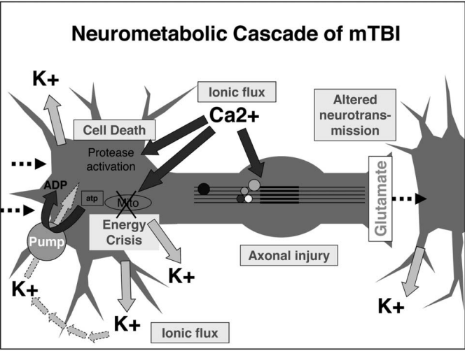
After an injury, an ionic flux happens within the brain. This means that the ion channels are not working to maintain a proper membrane potential within neural cells. A spread of inhibition is produced from the site of injury referred to as “spreading depression.” In an effort to reinstate homeostasis, ion channels that require energy become overworked. Intracellular energy is depleted, cells enter hyper glycolysis, and the brain shifts into an energy crisis. Neural cells use energy up before more ATP can be synthesized. Free radicals are produced, and metabolic pathways are continuously shifting for a 7–10-day period [1]. This is when the brain is most vulnerable to repeated injuries.
Structural damage is also a concern. Recent studies have demonstrated cytoskeletal anchor points in the brain to be a primary target for neural injuries. Axons, especially unmyelinated, are more vulnerable to these types of injuries as well. Disruption with cytoskeletal components can lead to axonal disconnection in which neuronal axon connections are severed. In some cases, axonal disconnection is the result of atrophy or neuron shrinkage. Chronic axonal dysfunction can result in impaired cognitive function and slowed reaction time.
Changes with the glutamate NMDA receptor can lead to alterations in normal developmental plasticity, electrophysiology, and memory. These changes can also lead to immediate early gene activation and different calcium flux patterns. E/I balance is altered due to changes in inhibitory transmission with GABA and its receptors. Inflammation is caused by an increased activation of microglia and an upregulation of cytokines and inflammatory genes.
Effects of Repeated Injury
There seems to be more extensive impairments following repeated mild TBI. There is little cell death observed after one mild TBI, but following a single moderate or severe TBI, cerebral and hippocampal degeneration and a loss of dopaminergic neurons in the substantia nigra is observed. DTI and MRI may be able to detect damage to cerebral networks, slowed conduction, and deficits in neurotransmission. DTI is better at detecting injuries of white matter by measuring where water diffusion is happening and in what direction within the brain. This measurement is referred to as fractional anisotropy [1].
The alterations that take place after an injury lead to the hallmark symptoms of concussions. Because of the energy crisis, the period after the injury and before the crisis subsides, the brain is especially vulnerable to repeated injuries. This can be what makes the healing process difficult to navigate. Since everyone’s situation is different, not allowing enough time for the brain to heal before returning to activity is common. This is dangerous because repeated injuries can lead to chronic traumatic encephalopathy, a condition involving aggregated tau proteins. These proteins are hallmarks of neurodegenerative diseases such as Alzheimer’s.
Allowing time for the brain to heal is the best treatment option to date. This poses an issue for athletes who are eager to return to play as soon as possible. It is important that people become educated on the risks of concussions and TBI and how they can lead to serious long-lasting effects and dysfunction if the proper precautions are not taken.
References
- Giza, C. C., & Hovda, D. A. (2014). The New Neurometabolic Cascade of Concussion. Neurosurgery, 75(Supplement 4), S24–S33. https://doi.org/10.1227/NEU.0000000000000505