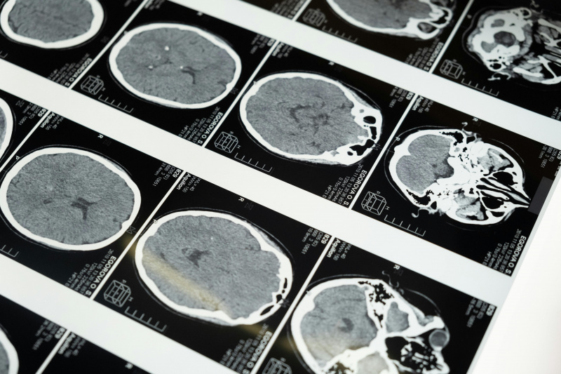
Concussions and Neuroinflammation: What Happens After Impact
When someone experiences a concussion, also known as a mild traumatic brain injury (mTBI), the damage is not always apparent through symptoms. Instead, much of the injury occurs on a chemical level inside the brain. After the initial impact, researchers have observed multiple pathologies to TBIs, including ionic flux, glutamate release, energy crisis, cytoskeletal damage, axonal dysfunction, and altered neurotransmission.[1] To read more about these processes, click here. One of the most important pathologies involved is neuroinflammation.
Many people assume inflammation in the brain only happens when the blood-brain barrier is damaged, allowing harmful substances to leak in. However, neuroinflammation can occur independently of changes in blood-brain barrier permeability. Brain cells, such as microglia and astrocytes, can trigger an inflammatory response and secrete cytokines on their own. These cells become activated after a concussion and invoke an immune response in the brain, including neuroinflammation.[2]
The Brain’s Inflammatory Response After a Concussion:
Inflammation following a mild traumatic brain injury can be either beneficial or detrimental, depending on how long it lasts and how intense it becomes. In the early stages, inflammation can help protect neurons and support recovery. Problems arise when this response becomes excessive or prolonged.
Several cytokines play major roles in this process:[3]
Interleukin-1 (IL-1) is part of a family of cytokines, although the most important forms are IL-1α and IL-1β. IL-1α spikes immediately after a concussion, while IL-1β increases gradually over several days. The levels of these cytokines depend on the severity of the trauma. IL-1β also stimulates the release of other pro-inflammatory molecules, including tumor necrosis factor-alpha. When IL-1 is hypersecreted, it can create a toxic inflammatory environment that can result in cell death in severe cases.
Tumor Necrosis Factor-Alpha (TNF-α) rises rapidly and usually returns to normal within 24 hours of the initial injury. TNF-α can also be protective or harmful. Its response depends on which receptor it binds to. Binding to the p55 receptor is associated with pathological effects, while binding to the p75 receptor supports neuroprotection.
Interleukin-6 (IL-6) has both pro- and anti-inflammatory roles and is stimulated by TNF-α. High levels of IL-6 have been detected for weeks after severe injury. One benefit of IL-6 is that it increases the production of nerve growth factor in astrocytes, which helps suppress TNF-α and IL-1β. This suppression keeps levels from rising too high and causing neurotoxicity.
Transforming Growth Factor-β (TGF-β) is an anti-inflammatory cytokine that peaks within 24 hours of injury and promotes tissue repair by suppressing inflammation. However, excessive levels can interfere with the brain’s own repair mechanisms and increase vulnerability to infection.
If you are interested in reading more about the involvement and effects of these cytokines in neuroinflammation, click here.
Why This Matters:
Although neuroinflammation can be harmful, it is also essential for neuronal growth and recovery after concussion and can even be used as a promising treatment option. Understanding the timing and interaction of these cytokines suggests that future treatments may focus on carefully timed combinations of pro- and anti-inflammatory molecules to reduce long-term neurological deficits after mTBI.[4]
[1] Giza and Hovda, “The New Neurometabolic Cascade of Concussion.”
[2] Patterson and Holahan, “Understanding the Neuroinflammatory Response Following Concussion to Develop Treatment Strategies.”
[3] Patterson and Holahan, “Understanding the Neuroinflammatory Response Following Concussion to Develop Treatment Strategies.”
[4] Patterson and Holahan, “Understanding the Neuroinflammatory Response Following Concussion to Develop Treatment Strategies.”