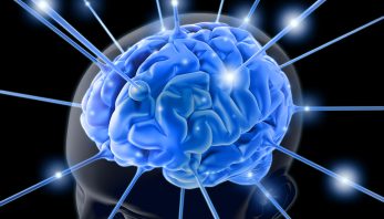
When I enrolled in neurochemistry, I had no idea what to expect from the course. I had no prior background in the subject matter besides an introductory biochemistry course. When coming into the course, I was expecting a normal lecture course where we would take notes, study, and eventually take a test on the material we learned. This was not the case for this course. In this course, we took a few weeks to study the basics of the brain covering basic receptors and signaling mechanisms. After we had this experience, we could finally get the full, unadulterated capstone experience.
From what I have learned from the various colleagues around the college, the capstone experience involves us taking a class in which we incorporate all of the material we have learned throughout our undergraduate career; a final hat on our learning experience. In neurochemistry, after our “basic training” session, we finally got to do this.
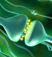
Each week we read through an article relating to a mental disorder such as Alzheimer’s disease, alcoholism, or even autism. We then took the time to study the pathways and their malfunctions in these diseases. Then, after we had read the article and discussed the “meat and potatoes” of the article, we had discussion every Friday morning about critical issues regarding the subject matter allowing us to apply our critical thinking skills we have learned throughout our undergraduate career. After these discussions, we would then compile our thoughts on the subject in a small, concise essay we would upload to a blog in which the world could read our thoughts.
Personally, I loved the way this class enveloped the capstone experience. We were able to back away from the stress of cramming for tests or writing a paper worth a significant portion of our grade and because of this, we were able to sit back, relax, and enjoy the material we were presented and relate it to critical issues that are faced throughout the world today. Neurochemistry also allowed us to use our previous education in chemistry and bring it all together in one class, which I believe is also a very important part of the capstone experience.
As well as encompassing the capstone experience, this course also embraced the colleges theme of B.R.E.W.; become responsibly engaged in the world. Along with our discussion of the science going along with the article, we also had very deep discussions regarding ethics of disease treatment and how we can apply our scientific knowledge to help the world. Because of this, we gained important experience in dealing with critical issues in the world and how we can approach them and “become responsibly engaged”.
All in all, I thought neurochemistry was a fantastic capstone course in chemistry. We were able to use the knowledge and skills we gained throughout our undergraduate career and apply it in sort of a “finale” course. By doing this, we also learned a bunch of new information about mental disorders that are very prevalent in the world today and how we as scientists are able to study them and treat or prevent them. In my opinion, the issues discussed in this course really embraced what Concordia tries to instill into its students; being able to think critically about issues in their area of study. I will embrace what I have learned in this class and the skills I have taken away and become more responsibly engaged in the world.
B.R.E.W.ing with Neurochemistry