Let’s talk about Obesity: a fast growing global epidemic

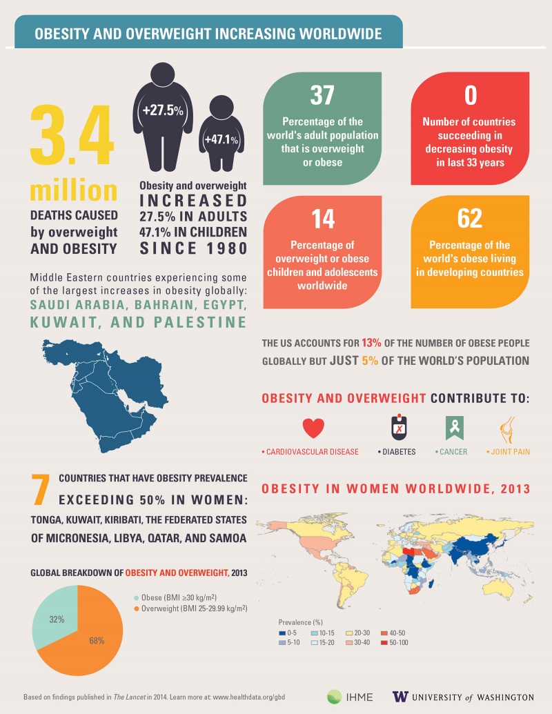
Obesity is now considered as a an epidemic that the public health is currently facing globally than malnutrition/ undernutrition. According to WHO in 2016, 2 billion adults were found overweight, of which 650 million of them are affected with obesity.

In the US, obesity has risen exponentially since the ’60s, which is also reflected with the busy “microwave” lifestyle that most Americans live, if looking at its prevalence in the United States. This thus implies a lack of time and investment in self-care, which in return opens the door for overconsumption of processed foods that are high calories, especially from fats and sugars.

This is very well alive in other parts of the world as well, regardless of the income level of the country. Below is a world map of obesity around the world.


Exposure to a high-fat diet and neuroinflammation in the Central nervous system has been shown to increase risks and causes for obesity, besides genetic predisposition. Changes in brain neurochemistry have impacts on the hypothalamus and its regulations for insulin and leptin levels and disrupt food consumption and energy.

This creates imbalance in food intake and energy consumption, caused by the insulin resistance that affect the hunger and satisfaction feelings.

The AgRP signals are released to create a feeling of hunger where as the alpha-MSH from POMC neurons allow for the feeling of satisfaction. The insulin resistance thus causes disruption, where there occurs more food intake than energy consumption. The insulin resistance can be caused by a high-fat diet or genetic mutation that cause neuroinflamation through the release of pro-inflamatory cytokines from the overactivity of mIcroglia and astrocytes.

ABT statement: A high fat diet is a measure trigger for obesity that is worth cutting down when considering underlying health issues, but there are various traditional cuisines around the world that have a fat heavy diet, therefore to what extent can culture be flexible to sustaining better lifestyle?

Artstract:


Insulin signaling and Obesity

What does insulin do

Insulin is a key regulator of adipose tissue lipolysis, which is the catabolic process leading to the breakdown of triglycerides stored in fat cells and release of fatty acids and glycerol (Langin et al., 2006).  Insulin regulates adipose tissue metabolism through direct effects on adipocytes (a cell specific for storing fat) and through signaling in the central nervous system by dampening sympathetic outflow to the adipose tissue (Shin et al., 2017). So, what about insulin in relation to POMC and AgRP?


Regulating Hunger

POMC and AgRP neurons

The insulin receptor is expressed in both AgRP and POMC neurons. Insulin can both inhibit AgRP and POMC through hyperpolarization or can activate these neurons as a result of heterogeneity (Shin et al., 2017). Insulin and leptin act directly on neuronal subsets in the ARC of the hypothalamus to control energy homeostasis. POMC neurons are activated, while AgRP neurons are inhibited. Then the MC4R-expressing neurons in the PVN. If one is fasting, the AgRP levels are increased and the POMC levels are reduced which leads to decreased MC4R signaling. When one has eaten, AgRP levels are diminished and POMC levels increase, which triggers MC4R signaling, stimulating energy expenditure (Jais et al., 2017).


Insulin resistance in Obesity and Type 2 Diabetes


Insulin resistance in Obesity and Type 2 Diabetes is classified by decreased insulin-stimulated glucose transport and metabolism in adipocytes and skeletal muscle and by impaired suppression of hepatic glucose output. In this situation, insulin binding, receptor phosphorylation, and phosphorylation of IRSs are reduced. In general, this means that PI3K activity associated with both IRSs is impaired (Langin et al 2000).


Why is this Important?

Figure 1: https://www.cdc.gov/obesity/data/prevalence-maps.html#overall

According to the CDC, the US obesity prevalence was 42.4% in 2017-2018. Obesity can lead to other disorders including, heart disease, stroke, type 2 diabetes and certain types of cancer. These are among the leading causes of preventable, premature death (CDC et al., 2021).


What can I do to prevent this?

Healthy Plate

There isn’t necessarily a diet you can do to help prevent insulin resistance in your body, you must be willing to change your lifestyle all together. Obviously, everybody knows you should eat healthy, this isn’t new information, but it is important that when eating unhealthily it is in moderation. Although you should not skip meals because this causes your insulin and blood sugar levels to go up and down leading to more body fat (Moore et al., 2019). Try your best to adopt healthy habits slowly and they will become permanent.


Works Cited


Centers for Disease Control and Prevention. (2021, September 30). Adult obesity facts. Centers for Disease Control and Prevention. Retrieved November 22, 2021, from https://www.cdc.gov/obesity/data/adult.html.


Jais, A., & Brüning, J. C. (2017). Hypothalamic inflammation in obesity and metabolic disease. Journal of Clinical Investigation, 127(1), 24–32. https://doi.org/10.1172/jci88878

Kahn, B. B., & Flier, J. S. (2000). Obesity and insulin resistance. Journal of Clinical Investigation, 106(4), 473–481. https://doi.org/10.1172/jci10842


Langin, D. (2006). Adipose tissue lipolysis as a metabolic pathway to define pharmacological strategies against obesity and the metabolic syndrome. Pharmacological Research, 53(6), 482–491. https://doi.org/10.1016/j.phrs.2006.03.009


Moore, W. (n.d.). Insulin resistance diet: How to use Diet to prevent diabetes. WebMD. Retrieved September 23, 2019, from https://www.webmd.com/diabetes/diabetes-insulin-resistance-diet.


Shin, A. C., Filatova, N., Lindtner, C., Chi, T., Degann, S., Oberlin, D., & Buettner, C. (2017). Insulin receptor signaling in POMC, but not agrp, neurons controls adipose tissue insulin action. Diabetes, 66(6), 1560–1571. https://doi.org/10.2337/db16-1238


Obesity and the Endocannabinoid System

Obesity Basics 

Figure 1: https://www.cdc.gov/obesity/data/prevalence-maps.html#overall

Obesity has been identified a top 10 global health problem by the World Health Organization. According to the CDC, nearly 42.4% of the adult population in the US were obese in the year of 2017. In figure 1, the color of each state represents the percentage of obese individuals in that state within 2020.

An important contributor is increased caloric intake without increased physical activity. An increase in calorie consumption is conducive to an increase in appetite from increasing craving and liking of food. The main brain system involved in craving and liking is the endocannabinoid system (ECS).

The Endocannabinoid System

The ECS functions to adjust behavior and metabolism according to food availability. Its action range from regulating sensory responses to developing preference for the consumption of high caloric food. In today’s world it appears that the ECS favors obesity and metabolic disease as food is readily available, especially “junk” food.

The ECS receptor in the brain is the cannabinoid-1 receptor (CB1). Researchers have examined the effects of activating and deactivating this receptor has on the body.

CB1

Cannabinoids and endocannabinoids bind this receptor. Figure 2 represents a simple schematic of the retrograde signaling of the ECS system.

Figure 2: A basic representation of ECS signalling in neurons. https://gaiaguru.co.uk/cannabinoid-receptor-1-basics/

Cannabis has been documented as promoting eating through strong cravings and an intensification of the sensory and hedonic properties of food. It has been demonstrated that the hyperphagic actions of THC (one of the psychoactive constituents of cannabis) were mediated in large part by cannabinoid CB1 receptors. Additionally, researchers saw that, systemic anandamide (an endocannabinoid) administration caused an increase in food intake. Later experiments confirmed that other endocannabinoids (2-AG and noladin ether) also increased food intake. Therefore CB1 agonists promote eating behaviors that lead to obesity.

In addition to CB1 agonists, researchers determined that CB1 antagonist suppressed food intake. They found that mice without CB1 receptors had a reduced sensitivity to the motivating properties of food, exhibiting reduced rates of responding for food and lower breakpoints when given a task to obtain food. In effect, the stimulatory actions of cannabinoids on eating resembles the changes that occur in food deprivation: levels of

Figure 3: CB1 receptor activation induces inhibitory effects on insulin signaling. doi:10.3390/ijms20092109 

anandamide and 2-AG increase after fasting. Therefore, this shows that endocannabinoids have an effect on the food intake of the brain without any drug influence.

In figure 3, you can see how some researchers believe CB1 signaling can affect insulin signaling that could in turn mediate weight gain. This activation of the CB1 signaling can be done by endocannabinoids (anandamide) or cannabinoids (THC).

Figure 4: doi:10.3390/ijms20092109

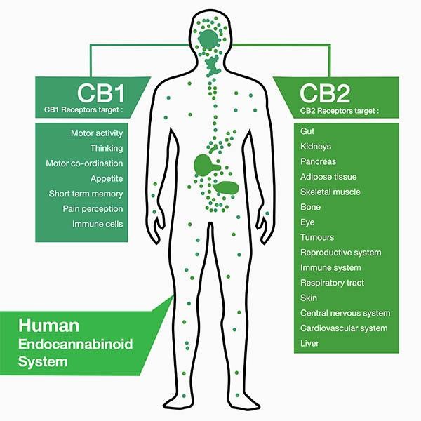
In figure 4, it describes the different locations of CB1 receptors and their pathological implications. As you can see the ECS is implicated in many agents of obesity development.

Overall, CB1 agonists show to increase wanting and liking of food AND CB1 antagonists show to decrease these. The endocannabinoid system plays an important role in the development and control of obesity. However, the exact mechanisms of these neurotransmitters on a cellular level are still not completely understood. Therefore, research must continue to clarify how endocannabinoids and cannabinoids can mediate obesity and how they might be able to be a part of treatment.

References:

https://www.cell.com/trends/endocrinology-metabolism/fulltext/S1043-2760(15)00140-X

https://www.mdpi.com/1422-0067/20/9/2109

https://gaiaguru.co.uk/cannabinoid-receptor-1-basics/

https://www.cdc.gov/obesity/data/prevalence-maps.html#overall 

https://doi.org/10.3389/fnins.2017.00293 

How You and I Can Help Keep People Safe in Contact Sports

Concussions are no joke, and it’s nice to see that people are finally beginning to take them seriously both on and off the field. The most dangerous part of a concussions is the chance of re injury, which means the most important piece to helping people with concussions is to stop them from a secondary impact. This is usually done by stopping players from playing for a while, but in order to do that the first step is recognizing the problem, which can be a challenge for many reasons. Such reasons include wanting to keep playing, dismissing it as a headache, or even ignoring the concussion to keep a player on the field. As concussions have become more recognized and dangerous, players and coaches need to change that mentality to one of a fast recovery. One of the best ways to get that first recognition and fast recovery in is by using the SCAT test.

The SCAT test is designed to be both user friendly and efficient, combining two of the key problematic factors regarding concussion diagnosis. Every coach, family member, and player should be able to use this tool effectively due to the functional layout and simplified instructions. By having these tools accessible to the public, concussions can be traded much more efficiently provided they are given the attention they deserve.

Below is an image of the first page of a SCAT-3 test as it would be administered in the field. One of the main strengths of the test besides its ease of use is the ability it has to cover all its bases. The test begins with important baseline information, such as level of consciousness and basic cognition, which are the most important indicators of a severe concussion. From there the test moves to a self-report, which can be both helpful and harmful depending on how it’s used. If this test were stand-alone it could lead to a series of issues, but the SCAT test instead uses it as a part of the baseline establishment, which allows for much more important information to be gathered by the observer. The test then goes into an in depth analysis of how the test taker performs on a variety of physical and cognitive tasks, with a score value assigned to each test. It may seem like a lot to the casual observer, but the level of detail combined with the ease of administration makes these tasks both manageable and effective at diagnosing a concussion. After the tests are done a concussion diagnosis can either be made or ruled out, and if a concussion is determined to exist the test outlines the next steps to take for seeking help.

Source For SCAT Test

There is still much work to be done regarding concussions and public opinion, but focusing on catching them before they can cause permanent damage is an important first step in recognizing the severity of concussions and moving towards a safer recovery period for people that live in high contact potential scenarios like contact sports or manual labor. In conclusion, ask your local coaches if they would be willing to have a copy of the newest SCAT test on hand, they never know how important it could be for their team!

Source for Featured Image

Concussion & Behavioral Changes

About 1.6 to 3.8 million sports and recreational concussions occur in the USA each year. 1 in 5 of these individuals will develop mental health concerns and disorders. Signs and symptoms that are severe can last as long as 6 months. Females, the elderly population, athletes, and individuals in the military are the most susceptible to concussions. Females are more susceptible because of weaker and thinner neck muscle fibers. Below is an image showing these differences: (3, 5)

Mental Health symptoms after experiencing a concussion include but are not limited to:

  • social anxiety
  • irritability
  • anger
  • depression
  • feelings of overwhelm
  • general anxiety
  • mood swings
  • emotional lability
  • impatience
  • difficulty concentrating
  • brain fog
  • sensitivity to light
  • fatigue or burnout
  • memory loss
  • sleep problems

Researchers have stressed that an individual’s personality doesn’t change after a concussion or traumatic brain injury but their behavior does. Their mood can be altered due to the symptoms of a TBI such as poor quality or lack of sleep, the frustration of not remembering things, feeling burnout easily, developing anxiety, etc. Personality is described to be a part of the person such as their dislikes, favorite food, activities, color, people, etc. How the individual reacts to such variables may change but they are not necessarily gone. This is not to say that personality can’t change because it can be depending on the location of the injury such as Phineas Gage in which his personality and behavior dramatically changed after suffering an injury to his frontal lobe. The diagram below shows the impact of the incident on the damaged regions.  (7, 1)

One study found that experiencing a TBI can increase your chances of developing a mental health disorder by 439%. Many people suffer from social anxiety, irritability, anger, depression, feelings of being overwhelmed, general anxiety, mood swings, or emotional lability (uncontrollable or inappropriate crying) after their injury. (3)

Concussions disrupt the communication between neurons and the blood vessels along with damage white matter within the brain. The damaged pathways will take a couple of weeks to heal but full recovery is expected for nonrepeated or infrequent concussions. In 30% of individuals who experienced concussions noticed a long-lasting slow and disruptive cognitive processing and responses. (1)

Females, the elderly population, and individuals who have had prior experiences of concussions, seizures, migraines, learning, mood, and anxiety disorders are the most susceptible to experiencing PCS (post-concussion syndrome). PCS is when the individual has prolonged symptoms and has continued experiencing such signs and symptoms past the expected recovery time given by doctors. The NINDS found that 20% of individuals who were in the military had experienced injuries during Iraq and Afghanistan. Of this 20%, 83% obtained a concussion or mild traumatic brain injury. Depression after sustaining a concussion is a common symptom. These individuals are twice as likely compared to individuals without TBI’s, to be at higher risk for self-harm and attempting suicide. Depression may contribute to the struggles that come with social interactions after a concussion as the person becomes easily fatigued and irritable due to intense lighting, loud noises, or too much movement and stimuli. (5)

Overall, concussions can lead to behavioral changes due to the symptoms and signs followed by the incident. Seeking professional help on how to manage these symptoms and signs is encouraged.


Resources:

  1. https://www.cognitivefxusa.com/blog/personality-changes-after-a-brain-injury-or-concussion
  2. https://www.nih.gov/news-events/news-releases/mental-health-disorders-common-following-mild-head-injury
  3. https://sciencenordic.com/biology-denmark-depression/head-injury-can-cause-mental-illness/1395035
  4. https://www.cognitivefxusa.com/blog/multiple-concussions-effects-and-treatment
  5. https://www.medicalnewstoday.com/articles/326227#complications
  6. https://www.spokesman.com/stories/2019/apr/03/researcher-mental-health-issues-often-progress-aft/
  7. https://www.npr.org/sections/health-shots/2017/05/21/528966102/why-brain-scientists-are-still-obsessed-with-the-curious-case-of-phineas-gage

Protection of the most important organ from concussions, the Brain

We all want to keep our friends and family safe. That includes protecting them from outside harm and making sure that they won’t get hurt. The problem is that it cannot always be prevented. One example of an adolescent child getting hurt is through a concussion and it is even more common to occur if that child is in sporting events. A concussion is described to be a traumatic brain injury (TBI) resulting from a blow to the head causing the brain to move back and forth at a fast pace [1]. This rapid and sudden movement causes the brain to form chemical changes and sometimes damage important brain cells [1]. A picture of what the brain is exposed to during a concussion is shown in the figure below. Concussions are potentially very dangerous as any damage to your brain can cause detrimental long term effects. 

A study on the neurometabolic cascade of concussion went into detail on what goes on in a person’s head when their brain experiences a concussion. Biomechanical injuries caused by a concussion lead to ionic flux and glutamate release triggering voltage gated ion channels [2]. This leads to the brain increasing ATP pumping to restore homeostasis, but in turn causes depletion of cellular energy reserves leading to multiple dysfunctions such as altered neurotransmission [2]. This shows a possible energy crisis involved after a concussion, possibly impairing other important mechanisms throughout the body. Because of this, important mechanisms may not be able to perform as adequately after the concussion symptoms have subsided. 

With these multiple changes happening in your brain, this can result in possible axonal injury causing potential damage to important sections. According to the same study on concussions, axonal injury causes disruption in transports resulting in impaired cognition, slowed processing, and slowed reaction times [2]. This implies that concussions can lead to impairment and possible destruction of neuronal receptors leading to become difficult in processing information and possible difficulty in motor behavior. This is scary to think about because this makes it possible that concussions could even cause paralysis. 

After a person experiences a concussion, there is a chance that post-concussive symptoms will stay. It should be known that while they are rare for a person who experiences one concussion, these symptoms can become more likely to occur in somebody that has had multiple concussions in a short period of time. Such as if they had repeated blows to the head in a football game. Some of these long term symptoms include but are not limited to headache, dizziness, memory problems, irritability, insomnia, depression, sensitivity to noise and light, and sometimes it can even cause a personality change [3]. With these effects in mind, it is no wonder why people say it is important to wear your bike helmet.

From these studies on concussions, people should be able to understand the dangers of concussions when doing a very physical activity that can involve the safety of your well being. The brain is the most important organ in your body and it is every person’s job to make sure it is protected from any outside harm it may come across. 

  1. https://www.cdc.gov/headsup/basics/concussion_whatis.html
  2. https://www.ncbi.nlm.nih.gov/pmc/articles/PMC4479139/ 
  3. https://www.healthline.com/health/post-concussion-syndrome#symptoms 

How does screen time negatively impact concussion recovery?

As technology is increasingly becoming a big part of our younger generation’s life, it is important to acknowledge its damaging potential in the long run. Though it can be seen as a helpful tool to keep them entertained in the midst of a busy time for the parents, there has been shown to be more downsides to the screen time than good The use of screens at the early development stage has not only been shown to cause development and neurological impairment ( such as in autism), but it also hinders the recovery process. This has been shown to have a negative impact on the recovery from concussion.

(Kohl’s building blocks)

A study was conducted on 125 patients aged 12 to 25, to test the effects of 48hr deprivation of screen time on recovery after concussive symptoms. Control had a shorter duration of symptoms (3.5 days) than 8 days for those allowed screen time right after their concussion.

The Post-Concussive Symptom Scale on a score of 3 points was used to assess a 22-symptom scale for the subjects in the study. Each symptom is  rated from 0 to 6 (severe).(Macnow & Curran, 2021)

(Middlebrook, 2016)

Dr. Mark Halsted, a pediatrician assistant at the University of Washington in St. Louis, has made remarks that ” It’s not just the prolonged sole exposure to screen time that can slow down recovery, but everything that involves high activity such as social events, school, etc.”

With exposure to video games, there is a high requirement for concentration and focus for prolonged time on cognitive expenditure.

  • Using screen has often shown to decrease in score for cognitive testing
  • Impairment in social and behavioral skills as seen in autism
  • Due to compromised white matter integrity, reduced cortical thickness as seen in different brain scans.
  • As you can’t keep straining an injured arm, a concussed brain should keep out of screens to avoid further injury

therefore, this information should not pass unnoticed because of how much screen time has become an intergrated part of our lifestyle. The same way you coulnd’t keep hitting a bruised arm, is the same way screen time and high demanding activities should be avoided for a concussed individual.

Works Cited

Kohl’s building blocks. (n.d.). Retrieved from Importance of Limiting Screen time: https://penfieldbuildingblocks.org/parenting-tips/importance-limiting-screen-time/

Macnow, T., & Curran, T. (2021). Effect of Screen Time on Recovery From Concussion. A randomized clinical trail. JAMA Pediatrics.

Middlebrook, H. (2016, October 21). New screen time rules for kids, by doctors . Retrieved from CNN: https://www.cnn.com/2016/10/21/health/screen-time-media-rules-children-aap/index.html


Necessary Action in Concussion Recovery

Artstract - Megan Blatti

Rest, Rest, and More Rest

Physical and mental rest are the primary recovery recommendations for those suffering from a concussion. Cognitive and physical rest

should include the avoidance of:

  • Screen time
  • Reading
  • Watching television
  • Listening to loud music
  • Any physical activity

Figure 1

It is shown that the majority of activities one should limit are primarily ones that involve cognitive function and stress. This is due to the energy crisis the brain undergoes when experiencing such trauma as a concussion. When the brain experiences trauma, it begins rapid attempts at restoring balance, or homeostasis. While doing this, ATP (energy) becomes depleted in efforts to recover. This energy depletion creates a slew of issues regarding signaling, neurotransmitters, etc. that may be mitigated via cognitive rest (Giza and Hovda, 2014). Although, it is not recommended that one rest during their entire recovery process (as in lying in bed in a dark room for multiple days). There have been cases in which such rest has shown to be detrimental to one’s recovery process. Complete rest, in which one avoids all stimuli, may instigate further rumination on one’s injury, creating depressive or anxious symptoms. Also, studies have shown that those prescribed with 5 days of rest experienced worsened post-concussive symptoms than those only prescribed 2 days of rest. So, although rest is necessary, it is only useful in moderation.

Pharmacological Intervention

Medication currently used to treat concussion are primarily intended to relieve concussive symptoms an individual is experiencing at that time. There are no FDA-approved medications for the treatment of concussion. Although, there are many pharmacological options one may pursue to lessen symptoms. The primary headache relievers include acetaminophen and NSAIDs, and amitriptyline and nortriptyline. The former pain relievers have actually been shown to be overused in those with concussions, and have the possibility of worsening symptoms. The latter medications are tricyclic antidepressants that may be used for migraines. They have been shown to relieve headache symptoms in pediatric concussions. Stimulants, such as amantadine and methylphenidate, may both be used to mitigate cognitive fatigue. Amantadine is a dopamine agonist that may theoretically improve brain functioning, while methylphenidate has been shown to improve cognition and mental fatigue after TBI in adults. In order to improve sleep in those with concussions, melatonin and trazodone are both recommended sleep aids. Trazodone is a serotonin antagonist often used for insomnia. Lastly, hypertonic saline may be used in severe TBI to reduce intracranial pressure. Also intravenous migraine therapy, such as metoclopramide or prochlorperazine, have been shown to significantly reduce pain in 93% of patients (Halstead, 2016).

Figure 2

Avoidance of Subsequent Re-Injury

The primary action one can take to ensure full recovery from a concussion is avoidance of a repeat concussion. This is especially important considering that it has been shown individuals are more susceptible to secondary injury following a primary TBI. This risk for subsequent injury is highest within the first 10 days of the initial trauma. This is likely to biological vulnerability from the ongoing energy crisis in the brain, as discussed previously. Magnetic resonance spectroscopy has displayed that reductions in a specific metabolite, NAA, took 15 days longer to return to normal levels in those with a repeat concussion, versus those with only a primary injury. Studies have also shown that repeat mild TBI may result in white matter damage in the brain, as well as cognitive impairments. If one experiences a second concussion before full recovery from the initial one, it is likely that their symptoms will worsen and last much longer. Although, if one experiences a repeat concussion after full recovery, it has been seen that these act like two, separate injuries (Giza and Hovda, 2014).

References

Giza CC, Hovda DA. The New Neurometabolic Cascade of Concussion. Neurosurgery. 2014; 75. doi:10.1227/neu.0000000000000505

Halstead ME. Pharmacologic Therapies for Pediatric Concussions. Sports Health: A Multidisciplinary Approach. 2016; 8(1):50–52. doi:10.1177/1941738115622158


Concussions leading to CTE

The Basics

Figure 1: https://www.mdpi.com/2411-5142/4/2/37

A concussion is a condition of neurological signs and symptoms that are caused by a biomechanical force to the brain. Symptoms of a concussion include headache, nausea or vomiting, dizziness, poor balance, light/noise sensitivity, fatigue, memory problems, confusion, etc. The cause of a concussion is a bump, blow, or jolt to the head or body that causes the head to move rapidly back and forth, as seen in Figure 1. The sudden movement of the head causes the brain to “bounce” around or twist in the skull causing injuries to the brain. The injuries to neural tissue caused by a concussion are predominantly functional or microstructural. Functional injuries include ionic shifts, metabolic changes, and impaired neurotransmission. Microstructural injuries include axonal stretch, axonal disconnection, inflammation, cell death, and cytoskeletal damage.

Concussions & Sports

Figure 2: https://www.vox.com/science-and-health/2018/2/2/16956440/super-bowl-2020-concussion-symptoms-cte-football-nfl-brain-damage-youth

Between 1.7 to 3 million sport-related concussions happen each year with around 300,000 of those from American football. About 5 in 10 concussions gounreported or undetected. According to a study published in 2005, there are about 0.41 concussions per NFL game with 67.7% of concussions involving impact by another player’s helmet. 92% of players who sustain a concussion return to practice in less than 7 days. This data is shocking; however, it is from 16 years ago. The technology to prevent concussions in football and the concussion protocol have changed. Nevertheless, the deadly costs of repetitive hits to the brain are being revealed.

Chronic Traumatic Encephalopathy (CTE)

CTE develops gradually from an accumulation of blows to the head or repeated concussions. Symptoms vary between individuals but tend to resemble other degenerative brain conditions like Alzheimer’s disease: memory loss, depression, mood swings, personality change, fatigue, anxiety, frustration, etc. There is no diagnostic test for CTE, thus, a diagnosis cannot be confirmed until the post-mortem brain can be examined. Football players have a high risk of developing CTE as they are subjected to repetitive head injuries.

From a study of 202 brains from deceased players of American football, CTE was diagnosed in 177 players (87%), including 110 of 111 former NFL players (99%). The most common cause of death for participants with mild or severe CTE pathology was suicide, 27% vs. 47%. 86% of former NFL players hadsevere CTE, compared to 56% of former college players and 56% of semiprofessional players. The level of neurofibrillary tangles correlated positively with the severity of CTE. Neurofibrillary tangles (NFTs) are abnormal accumulations of the hyperphosphorylated (too activated) protein

Figure 3: https://heavy.com/sports/2020/01/aaron-hernandez-cte-photos-brain/

tau. Normally, tau supports the structure of the neuron by stabilizing microtubules which help guide nutrients and molecules down the cell. Within tangles, hyperphosphorylated tau detaches from the microtubules and stick to each other, forming tangles that block the neuron’s transport system from inside the cell. In figure 2 you can see the mass amounts of NFTs in a region of Aaron Hernandez’s brain. Additionally, there were deposits of amyloid- in all stages of CTE. This protein forms plagues that disrupt communication between neurons.

NFTs and amyloid-  plaques are key markers of Alzheimer’s disease and associated with symptoms of impulsivity, depression, apathy, anxiety, explosivity, aggression, memory loss, and confusion. Among those that showed CTE pathology, 42.3% presented with cognitive symptoms in life, 43.2% showed behavior/mood symptoms, and 14.4% presented with both cognitive symptoms and behavior/mood symptoms.

Aaron Hernandez

A particular case study of CTE in a football player showed severe CTE pathology and the symptomology described with severe CTE. This case is of Aaron Hernandez.

Aaron Hernandez was a tight end for the New England Patriots until his career ended with his arrest for the murder of Odin Lloyd. Two years after his conviction for first degree murder, Hernandez committed suicide within his prison cell. Pre-mortem, professionals thought he had CTE, but this was confirmed after his death when

Figure 4: https://heavy.com/sports/2020/01/aaron-hernandez-cte-photos-brain/

researchers were allowed to examine his brain. For a little bit of background Hernandez had been playing football since high school. He had many of the symptomology of CTE: changes in mood, aggression, depression, mood swings, personality changes, and memory loss. His brain: through Figure 3 you can see there is an atrophy of his brain specifically in the fornix and hippocampalregions. They also saw perforations of the septum pellucid and deposits of tau that you can see on this picture. Also, you can see that he had a lot of ventricular enlargements.

The researchers who examined Hernandez’s brain claim that it is the worst case of CTE they have ever seen in someone of his age of 27 years old.

After the death of Hernandez, his family filed a lawsuit against the New England Patriots and the NFL for being fully aware of the damage that could be inflicted from inflicted from repetitive impact injuries and not disclosing, treating, or protecting Hernandez from those dangers. They have since dropped this charge.

CTE makes a murderer?

I’m not sure that a neurodegenerative disease can make a murderer. In my opinion, neuronal dysfunction can be an aggravating trigger for homicidal tendencies. But this does not mean having CTE excuses such heinous acts. Instead, I believe CTE is a serious disease that is experienced by the many who experience repetitive traumatic brain injuries and shows itself as a change in key brain functions. CTE is something that should put fear in athletes, coaches, parents, and society. The development of CTE changes the brain and changes the person. And the truth is staring us in the face, if individuals continue to hit their head whether it be in combat, self-injury, accidents, or football they may see a change in themselves only explainable by CTE.

Sources:

https://www.nhs.uk/conditions/chronic-traumatic-encephalopathy/

https://heavy.com/sports/2020/01/aaron-hernandez-cte-photos-brain/

https://www.ncbi.nlm.nih.gov/pmc/articles/PMC6916416/

https://www.ncbi.nlm.nih.gov/pmc/articles/PMC3624697/

https://jamanetwork.com/journals/jama/fullarticle/2645104 

Does Fat Make You Fat?

When addressing the role of the high fat diet and obesity, it is important to evaluate the types of fats in a given diet. While a high fat diet can lead to increase in obesity, a low fat diets as intervention for obesity is not supported. Rather, increasing the intake of unsaturated fatty acids over saturated fatty acids, carbohydrates, and proteins, can be beneficial for the obese population. Therefore, one must evaluate the types of fat intake that one is receiving, and aim to increase and supplement polyunsaturated fatty acids and some monounsaturated fatty acids when reversing obesity and insulin resistance.

So, what are the types of fat? Which types are “good?”

Trans fat is a byproduct of hydrogenation that is used to turn healthy oils into solids to prevent from becoming rancid. It has no known health benefits and is banned in the United States. It is known to increase the amount of LDL cholesterol in the bloodstream, while reducing the good, HDL, cholesterol. It also causes massive inflammation, and contribute to insulin resistance.

Saturated fats are solid at room temperature. They are found in red meats, whole milk, cheese, coconut oil, and baked goods, etc. A diet that is rich in saturated fats can increase total cholesterol.

Monounsaturated and polyunsaturated fats are good fats that come from vegetables, fish, nuts and seeds. In particular, monounsaturated fats have a single carbon to carbon double bond. Foods such as olive oil, avocados, nuts, and foods familiar to the “Mediterranean diet” are high in monounsaturated fats. Polyunsaturated fats are essential fats. They are required for normal body functions and humans need to obtain them from food sources. They have two or more double bonds. They are used for many different processes in the body, mainly for building cell membranes, cover our nerves, and are used in blood clotting, muscle movement, and controlling inflammation. Eating polyunsaturated fats instead of saturated fats or highly refined carbohydrates reduces LDL cholesterol and lowers triglycerides. The two main types of polyunsaturated fats are omega-3 and omega-6 fatty acids.

Omega-3 fatty acids can be found in salmon, flaxseeds, walnuts, and mackerel, shrimp, hemp and chia seeds, among others. Omega-3 fatty acids are known to prevent and treat heart disease and stroke, they reduce blood pressure, raise HDL, lower triglycerides, and restore leptin and insulin sensitivity, especially in the obese population. The figure below shows how omega-3 PUFAs protect cell membranes through exerting anti-inflammatory effects through modulating NF-kB signaling, NLRP3 inflammasome, PPARa/y, GPR120, and TGF-B signaling. Omega-6 food sources include vegetable and corn oils, sunflower seeds, peanut butter, eggs, and almonds, and they have been linked to protect against heart disease.

The ratio of omega-3 to omega-6 fats is important, especially in regards to inflammation and it is recommended that rather than decreasing the amount of omega-6s in your diet, which can be anti-inflammatory, it is better to increase your omega-3 intake.

Overall, while some fats are harmful to our health and bodily functions, and contribute to diseases like obesity and insulin resistance, high quality fats like polyunsaturated fatty acids can actually protect and reverse obesity and insulin resistance. 


Spam prevention powered by Akismet