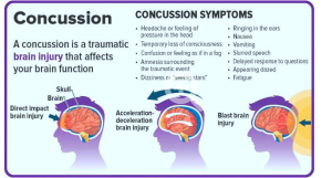
Figure 1: A mapping of general symptoms associated with concussions.
What is a concussion?
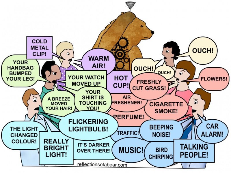
Put simply, a concussion is the result of a mild traumatic brain injury (mTBI). This is common when the brain bounces off of the inside of the skull because of a great force that is applied to the head in a short amount of time. Common examples come from collisions in sports like football and hockey as well as head injuries resulting from falling or car crashes. The cellular mechanics of these events are pictured below in Figure 2.
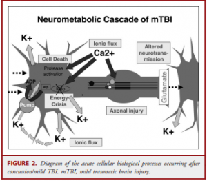
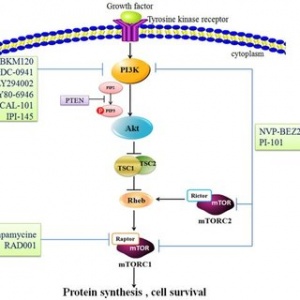
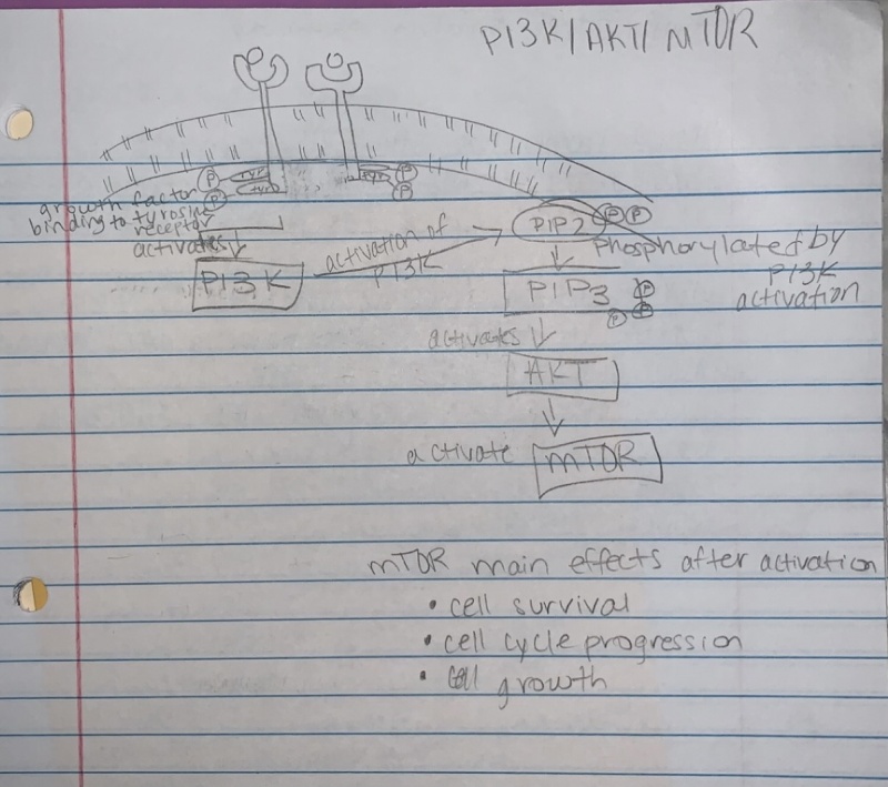
Figure 2: A map of what happens on a cellular level when the brain experiences a TBI.
To explain what is happening in Figure 2, it makes to try to follow a chronological timeline after injury. First, because of the injury, holes in the plasma membrane are going to develop. This leads to an ion flux because the axon is no longer to maintain an ion gradient between the inside and outside of the cell. Because of this, an energy crisis is going to occur for 2 main reasons: ion pumps and mitochondria dysfunction. Ion pumps are going to be overworked trying to maintain an ion gradient (which will be nearly impossible because of the holes in the plasma membrane due to the injury), which is important because these are ATP activated channels. So, a lot of energy is going to be used attempting this process. To further propagate this energy crisis, the cell becomes overwhelmed by calcium ions, which it will then deposit in the mitochondria for storage. If there is too much of this calcium deposition, the mitochondria will stop working. This effectively shuts down ATP production by the mitochondria. Instead, the cell must now use glycolysis to produce ATP, which is much less efficient. Overall these events lead to a stressed out cell.
Figure 3: Logo from the ImPACT testing website.
The importance of knowing recovery time
With neural damage as serious as mentioned above, it is very important for people to know when they have a concussion. Subsequent injury during a healing period from an initial mTBI can cause very serious brain damage and an associated longer recovery time. So what is there to test if someone has a concussion? Well one method is ImPACT testing. ImPACT testing is a way to test spatial memory and cognitive speed through a series of tests. It works because the user takes an initial baseline test when they aren’t experiencing a brain injury and then are tested against that baseline when there is a suspected brain injury. However, this method is subject to flawed results as the user could purposefully “tank” their first score so that they pass when they are experiencing a brain injury. The test itself is also subject to flaws. A user taking the test back to back might experience a very different score because of the nature of the test and the time component associate with it.
Figure 4: Picture of an MRI commonly used in fMRI and BOLD signaling analysis.
What to use instead?
Because of the flaws of the ImPACT test, I propose focusing on an emerging practice of using functional nuclear magnetic resonance imaging (fMRI) to analyze blood-oxygen level dependent (BOLD) signaling in the brain. BOLD signaling analysis works because deoxyhemoglobin (deoxygenated blood) is paramagnetic, causing it to appear on fMRI scans. So, these BOLD signaling studies can show where deoxyhemoglobin is present in the brain, which can be reasonably associated with oxyhemoglobin (oxygenated blood) and its deposition of oxygen in certain regions of the brain. Areas of the brain undergoing rapid repair (such as those that have experienced a mTBI) will need this oxygen. So, by using BOLD signal studies, we could determine a general recovery time associated with a certain level of mTBI experienced. This method is still in development because the association between deoxyhemoglobin and oxyhemoglobin isn’t as simple as I described above and this method is quite expensive.
The future of concussion recovery
By showing the severity of concussions to brain health, it is clear that more needs to be known about how concussion recovery occurs. Having a simply way to measure the pace of recovery, such as using ImPACT testing or fMRI BOLD signal studies, would greatly help. Perhaps there are other methods being developed like fMRI BOLD signal studies that give a more molecularly detailed description of recovery.

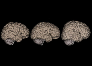
Figure 5: Artstract by Trenton Vogt. This figure depicts (very rudimentarily) how spatial memory can be impacted by a concussion.