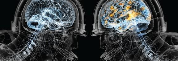
CAP-Ping Off My College Experience

The neurochemistry capstone is unlike any science class I have taken at Concordia. While the background of chemistry, biology, and neuroscience is helpful, it was learning how everything relates that was so integral to the articles. With each article came a new discussion, direction, and potential solutions to the many problems. It was great to have students with different backgrounds in the class which really helped our discussions throughout the weeks. We were able to challenge each other with new ideas or thought provoking questions.

    Each week, everyone in our class was able to contribute new information or a new perspective on the “article/disease of the week”. It was challenging to look at diseases from a multidisciplinary lens. We also stressed the importance of sharing science with the general population. While innovative research, conferences, and scientific literature is important, it is also important to communicate that information to the general public in understandable terms. Often times in society, this aspect is overlooked. We learned ways to be concise and explain things in a way that was educational and accurate. Communication in different mediums was a skill we utilized a lot in this class, with presentations, written essays, partnered discussions (or speed dating as we liked to call it), and blog posts. The practice of sharing scientific findings with the general public was especially achieved through the blog posts. We also were able to practice our artistic skills and abstract thinking to reduce complicated ideas into simple drawings or ‘ARTstracts’. Some of us who aren’t as artistically gifted found this to be a difficult component to the blogs!

Because many of the diseases result in degeneration and death, it elicits the need for a cure to be found. Before treatments can be discovered it is important to understand the pathogenesis and pathways that are disrupted during this disease states. Many of them are very complicated and have lots of difficult components and potential causes. We started the class with a foundation on some of the basic pathways involved in cellular metabolism and maintenance and then applied this knowledge each week.

            Week after week we were able to tackle difficult diseases with complicated pathways. We would dissect them, question them, investigate them, and discuss them. Our Friday discussions were always interesting, and usually took tangents with deep thought questions and social implications as well as application to our own lives. Being able to discuss the science, and culture of diseases with a group of fellow college students is quite rewarding. Sometimes I think we forget that most people do not wish to discuss the adenylyl cyclase pathway for 70 minutes on a Wednesday. Yet we plugged along tackling one problem at a time – often leaving with more questions than answers. The more we learned, the less we feel we know! While this concept is frustrating, it also leads to inspiration and innovation that I hope we are able to bring to the world one day.

We also split into teams and worked on a community action project which was another way to bridge the gap between science and the general public. My group looked at eating disorders, especially on campus and with athletes. By holding a talk on campus for students, coaches, and teachers we were able to reach an audience that normally would not hear about the role of neurochemistry in eating disorders. It is important to raise awareness, and keep people educated and informed as to what is going on in our bodies.

            I felt like this class was a good way to cap off my time at Concordia. It was a good culmination of many of the courses I have taken here at Concordia. I know I personally was able to connect Vaccinology, Anatomy and Physiology, Psychology 111 & 206, Immunology, Microbiology, Religion and Science, Organic Chemistry, Biochemistry, and Embryology with this class. On top of that my classmates had other classes and experiences to add to it! That just shows how important it is to have that encompassing experience that can connect many fields and ideas. The ‘liberal arts’ way of thinking is very important in approaching some of the issues we are going to face in the future. Having a wide basis of knowledge allows you to think more critically and connect dots between two seemingly unrelated fields.

As described in detail above this class has hit all the liberal arts goals: to instill a love for learning, develop foundational skills, develop an interdisciplinary understanding, examine culture, ethical, physical and spiritual self-understanding, and encourage responsible participation in the world.


ALS, Beyond the Ice Bucket Challenge

What is ALS?
Most people know amyotrophic laterals sclerosis (ALS) as Lou Gehrig’s disease after the professional baseball player who had to retire due to the illness. It was also the subject of a social media craze called the ‘ice bucket challenge” that aimed to raise awareness and funds for research. ALS is a progressive disorder that affects the motor neurons in the brain and spinal cord. The death of these neurons manifests as a gradual decline in voluntary movement that leads to paralysis and death.
Image result for ice bucket challenge
Individuals typically live 2-5 years after the first symptoms present themselves. The Center for Disease and Control estimates that roughly 10,000 to 15,000 people in the US are affected. Like Alzheimer’s, ALS is an age-related disorder with most cases occurring in individuals between the ages of 55 and 75.
Progression of Symptoms
-Early Stage

  • Trouble grasping small items
  • Changes in vocal pitch
  • Falling
  • Muscle cramps/twitches
  • Excess fatigue

-Middle Stage

  • Breathing becomes more difficult
  • Muscles tighten and become weak
  • Trouble walking and balancing
  • Eating and speech ability reduced

-Late Stage

  • Muscles become paralyzed
  • Speech is not possible
  • Breathing requires a ventilator

Two forms of ALS can occur, sporadic, and familial. The sporadic can impact anyone. This form accounts for up 90 to 95 percent of all cases. The familial form of ALS is the result of inheriting genetic mutations and is responsible for 5 to 10 percent of all cases.
The Latest Science
The cause of sporadic ALS is not well known. Scientists are working to understand the pathology to address the vital need of developing treatments and a cure. Recent research has implicated prolonged oxidative stress as a possible mechanism. It can lead to protein aggregation and therefore defective function of the proteins involved. It can also impact critical transport systems between the nucleus and cytoplasm of the cell by RNA binding proteins.
Oxidative stress can also cause mitochondrial damage by altering mitochondrial proteins. Through an unknown mechanism, these altered proteins can inflict damage on the mitochondria. These organelles are vital for the health of cells and damage can increase susceptibility of neuron death.
The conditions of oxidative stress and protein aggregation may also lead to improper protein metabolism. Protein dysmetabolism can negatively impact a big range of processes crucial for the survival and functioning of neurons.
It is worthy to note that familial cases of ALS are the result of mutations to genes that code for proteins involved in the pathways above.
Although we may be years away from a cure for ALS, promising advances are being made in understanding the molecular drivers of the disease. This will one day help develop effective therapies to increase the quality of life for those affected.
You Can Help!
I encourage you to visit the websites of organizations listed below to learn more and help fight ALS.

Image result for ALS


Parkinson’s Disease: What Is L-Dopa and How Does It Act?

Parkinson’s disease (PD) is a very tough disease to discuss because of its progressive nature and how debilitating it can be.  To any of you who knows someone that has PD you know exactly what I am talking about.  It starts out as barely noticeable tremors, stiffness, and moving slower.  Now the stiffness and slowing of movement can also be attributed to old age and that is what makes this disease so difficult to diagnose.  One other noticeable sign that only presents in Parkinson’s is the arms remaining stationary while an individual walks and also the inability to turn while walking without considerable time and effort.

In the United States there are about 1 million people that are diagnosed with the disease and around the world there are approximately 4 million people.  Every year there is about 60,000 people diagnosed with the disease in the United States alone.  Most PD onset is at the age of 60 or older, but as with many neurodegenerative diseases there is a small amount of early onset conditions.  This usually accounts for only 5-10% of all cases.
Parkinson’s disease is defined by an early selective loss of dopamine producing neurons in the substantia nigra and by the formation of Lewy bodies.  These Lewy bodies are an accumulation of alpha-synuclein and other misfolded proteins that cause oxidative stress and have other toxic effects that can eventually lead to dopamine neuron death.  Hence, the loss of some of some movements and the tremors that are commonly associated with the disease.  The reason alpha-synuclein aggregates in PD is due to it being phosphorylated by kinases that are dysfunctional.  In the normal human brain alpha-synuclein is phosphorylated only about 4% of the time, but in diseased brains this number can be upwards of 90%.  This causes the misfolded alpha-synuclein to aggregate and act like prions.  This simply means that the misfolded alpha-synuclein can bump into normal alpha-synuclein and cause that alpha-synuclein to become misfolded as well.  This leads to a continuous cascade of Lewy Body formation, which in turn leads to loss of dopamine producing neurons.

Now the classical treatment for Parkinson’s is L-Dopa.  The idea behind this drug is that there is simply a lack of dopamine in the brain so dopamine needs to be added to offset the loss of dopamine.  The main reason why dopamine is not given as a treatment is that it cannot cross the Blood Brain Barrier (BBB).  This is the barrier that prevents viruses, bacteria, and other molecules from entering the brain and causing problems.  However, the solution to this is giving L-Dopa that is an amino acid precursor as a treatment.  This molecule can cross the BBB and can be converted to dopamine once inside the brain.  The treatment of L-Dopa can alleviate the tremors and shakiness of Parkinson’s and also improve the ability to walk normally.  However, prolonged use of L-Dopa can lead to dyskinesia, which means involuntary muscle movement.

Parkinson’s disease is a progressive disease that eventually can lead to debilitating tremors, stiffness, and slow movement.  Like many other neurodegenerative diseases there is no cure.  However, L-Dopa can alleviate many of the symptoms and improve the quality of life of those diagnosed with the disease.  More research needs to be done to ensure that a cure is developed to stop this disease in its tracks.

The Stickiness of Parkinson’s Proteins

You have probably seen the muscle tremors that affect Parkinson’s patients.  Parkinson’s is a disease that kills dopamine-producing neurons in the brain.  While traits like intelligence, personality, and memory can decline, the most damage happens to the motor neurons that move our muscles.  Over a period of decades, muscle control gradually declines in Parkinson’s patients.  There are only a few treatments available for Parkinson’s, and they can only slow the progression of the disease or deal with the symptoms.

An innocent little phosphate

So what is actually killing the motor neurons?  The cause is still not completely clear, but we know that Lewy Bodies affect the neurons.  Lewy Bodies are clumps of sticky protein that build up in the neurons of people with Parkinson’s.  Lewy Bodies are also found in other forms of dementia.  They are made up of a protein called alpha-synuclein.  
And what makes this protein stick together?  The answer might be phosphate groups.  Phosphate groups are a molecule of phosphoric acid that gets added onto the protein.  For some reason, phosphate groups seem to make alpha-synuclein stick together.  Maybe it helps the proteins fit together like puzzle pieces.  You probably know phosphoric acid from the label on a can of pop.  Drinking phosphoric acid in soda is not dangerous (except to your teeth) because it takes special enzymes to add the phosphate groups onto proteins.  
Proteins that tack phosphate groups onto other proteins are known as kinases.  It turns out that most kinases are controlled by other kinases, so there is a complicated web of on and off switches inside our neurons.  If one kinase is mutated, it can affect dozens of other types of kinases, making them sluggish or hyperactive.  This can eventually lead to the clumps of neuron-killing protein.
Interestingly, when one piece of a-synuclein gets a phosphate group it encourages other ones to do the same.  This makes alpha-synuclein act like a prion disease in Parkinson’s patients.  The prion disease of mad cow disease made headlines when the public learned that their beef cows were eating the brains of other cows.  That’s a great way to spread a prion disease, and fortunately humans don’t normally swap much brain tissue.  But this does explain how muscle problems can start in an arm or leg and spread to the rest of the body, and how dementia can appear in late stages of the disease.  Parkinson’s can start in one area of the brain and slowly march across the brain from neuron to neuron.
The tremors of Parkinson’s make it impossible to eat and difficult to sleep.  People that work with their hands for a living are affected even more.  If we can stop proteins from sticking together in their neurons, we might be able to stop the disease.  Unfortunately, the root cause of the phosphorylation of a-synuclein does not just involve one gene.  It will take a lot of work by scientists and probably some supercomputers to put the puzzle pieces together so we can take other puzzle pieces apart.  
 

Concussion Background and Visual Imaging

In the good old days as some people would call it, if you get hit in football or any sport and you were knocked out it was almost seen as a sign of weakness.  No man went to the doctor to see what was actually going on in the brain, instead it was recommended to just “shake it off”.   However, with the bombshell report that the National Football League was hiding research about concussions and the potential lifelong effects that could result from multiple concussions, the research has become abundant.

Before discussing the research it is important to see the scope of how many concussions actually occur in the United States every year.  According to the Centers for Disease Control and Prevention (CDC) in 2013 about 2.8 million Americans were diagnosed with a Traumatic Brain Injury (TBI), most of them being mild TBI’s or as they are commonly known as concussions.  These TBI’s accounted for 50,000 deaths in the United States alone.  Among all the concussion diagnosed every year it is estimated that 70,000 people experience permanent damage to their brains. One interesting fact, is that the most common sport that causes concussions is not football, it is actually cycling.  However, football still experiences just over 36,000 annually.

As a result of a concussion many problems can arise.  Cytoskeletal damage can arise because the force of a concussion can cause the complex microstructural components formed by neurons and glia to be damaged. This damage to the cytoskeletal complex can also cause axon dysfunction.  This means that the axons on neurons can be overstretched causing them to become overly porous to excitatory molecules.  To the extreme end of this the axons can actually be snapped off at the axon hillock.  This is severe because this prevents axons from sending action potentials and communicating with the rest of the brain.  This can cause cognitive deficits and a lack of communication in the brain.  One other main contributor the symptoms of a concussion is increased inflammation.  The upregulation of cytokines and cytokine genes causes an increase in stress on neurons.  This increased stress if it is prolonged can ultimately lead to cell death and an overall loss of neurons.  Either way a concussion can be catastrophic, however what has arisen over the past several decades is the ability to see the effects of a concussion.

This is where the imaging arises. The first form of brain imaging is Diffusion tensor MRI (DT-MRI). It works by sensitizing the MRI signal to the random molecular motion of water molecules (diffusion) by addition of ‘diffusion encoding gradients’ to a standard MR pulse sequences.  Water molecules are more likely to travel down white matter (neuronal pathways) than they are to leave these pathways.  This allows you to visualize if any of these tracts are damaged, hence seeing if there is a concussion

The second method for visualizing TBI’s is functional magnetic resonance imaging or functional MRI (fMRI).  This works by using MRI technology that measures brain activity by detecting changes associated with blood flow. This technique relies on the fact that cerebral blood flow and neuronal activation are coupled. When an area of the brain is in use, blood flow to that region also increases.  So in essence decreased blood flow to a region of the brain would imply that there is damage to this area of the brain.

Based on the images above it is clear that concussions are not good for the brain and should be take very seriously.  If you or someone you know has a concussion make sure to see a doctor to plot a course of action and an effective treatment.

The Capstone Course Serves as a Sturdy Foundation for the Future

The Capstone Course Serves as a Sturdy Foundation for the Future
No project is complete without the finishing touch. A Christmas tree lacks luster without the star on top. A painting doesn’t shine without the varnish finish. A Concordia education is incomplete without a capstone course. As a chemistry major, the logical choice for my capstone class was neurochemistry, and I feel it was the perfect finishing touch to my Concordia career.
When I began my freshman year, I was set to be a chemistry major. I was good at chemistry and learning in lecture came easy. Neurochemistry was the first class I took that focused on independent learning, and it happened to be in my last semester at Concordia. I was initially nervous about the amount of biology at the beginning of the course but once we delved into the article analysis, it didn’t seem to matter as much.
While I was doing research at the University of Minnesota this past summer, the grad group I was part of had weekly “Lit Club” where they read current literature published that was similar to their research. I would sit through the meetings every Friday morning thinking that I would never be able to understand the articles they were reading. In neurochemistry, the articles were about the brain and signaling pathways of which I had no background knowledge. I learned that even though I lacked background knowledge, I was still able to understand the general message of each article on my own. It gave me hope that when I am part of a grad group in grad school, I will be able to understand the articles on my own and be able to contribute to the group discussion.
However, this is not the only reason this course has prepared me for the future. It has also taught me to enjoy reading articles of current research findings. Reading articles for class has seemed like a chore in the past, but for neurochemistry, I looked forward to reading about diseases and how altered brain functions resulted in signs and symptoms. I can apply this new enjoyment to my future by continually learning about new scientific discoveries.
As my time at Concordia comes to a close, I have looked back at everything I have learned along the way. Not only have I learned necessary chemistry knowledge, but I have now also learned how to apply my knowledge and continue learning on my own.
I truly believe that this capstone course was the perfect finishing touch on my Concordia education. Learning how to continue learning on your own, for scientists at least, I think is crucial to being a successful scientist. I look forward to my future and learning even more about subjects that I love.

Are Stress and Anxiety Hijacking Your Body?

Stress and Anxiety. Two words you hear often now a days. Especially if you are like me a college student. These two factors can negatively impact our lives and make life much more difficult. These two things are caused by our non-stop desire to be productive. People vary between how they deal with stress and anxiety differently and people differ in how easy they get stressed out or anxious. It’s weird how that is a widely accepted truth. But how does this occur? There are a couple of different reasons why.
A review article in the frontiers of psychiatry reveals that it is the formation of memories attributed to stress and anxiety. Although our bodies are designed in a way that you can make strong memories of stressful events so that in the future you can avoid them. When I say stressful event, the most likely stressor that a human would activate this mechanism would be a wild animal attacking them in the woods or something along the lines of that. But with the modern civilization and the way society is today we do not experience these sort of stressors often at all. This biological mechanism in the brain gets hijacked by the “stressors” we experience as humans almost everyday in the modern world. The paper describes that the adrenal gland releases corticotrophins, which are responsible for releasing adrenaline in the body and increase heart rate and alertness, which is characterized with stress and anxiety. In people with stress and anxiety disorders this becomes dysregulated and this very mechanisms attributes to the formation of the memories associated with the stressors.

In combination with increased corticotrophin release glutamate signaling pays into factor within stress related memories as well. Cellular mechanisms such as a dual histone activation of a protein complex called H3S10P-K14ac increases the production of proteins called c-Fos and Egr-1. These proteins are directly involved with forming strong memories and consolidation processes as well. This underlying mechanism can thus cause an increase of “noisy” thoughts that are characterized with the intense memories of the stressors, which can cause attention deficits. The Prefrontal cortex gets dysregulated as a result of this cascade because the connections between the hippocampus and the amygdala become desensitized.
There is a wide array of medications for anxiety, they include, Selective Serotonin Reuptake Inhibitors (SSRIs), Serotonin-Norephenipherine Reuptake Inhibitors (SNRIs), and Benzodiazepines. The mechanisms for SSRIs and SNRIs block the reuptake channels on neurons of these specific neurotransmitters that play important roles in mood stabilization. Recently it has been described that Histone modification of the phosphorylation and acetylation of the histone H3 complex (H3S10p-K14ac), leads the activation of gene transcription of IEG genes, which facilitate strong memory formation. This mechanism is strongly linked to anxiety disorders and it appears Lorezapam and well-known benzodiazepine blocks the phosphorylation and acetylation of the histone complex. Other anxiety medications and their mechanisms relating to these histone modifications are not well known. However, It has been found that the mechanism for benzodiazepines like lorezapram acts on GABAergic neurons. This relationship could possibly lead to the inhibition of the phosphorylation and acetylation of the histone complex. They are allosteric modulators of the GABA-A receptors. These receptors ligand-gated chloride channel and when activated hyperpolarize the target cell allowing Calcium ions to flow in. Benzodiazepines bind to these receptors in a specific pocket that induces a conformational change and allows GABA to bind.
It’s a terrible cycle. Stress and anxiety when in high doses can be maladaptive for everyone as indicated by this article. However, in this day and age these things are unavoidable. This is why developing a deeper understanding of these aspects and what they can do to a person is important. It is also important to manage these things so that the stress and anxiety levels to become too high where you can develop problems that can be very hard to deal with.

Understanding Concussions and Traumatic Brain Injury

If you were to ask a high school freshman what a concussion is, do you think they would be able to tell you? All I would’ve been able to tell you when I was a high school freshman is that a concussion is when you hit your head really hard. I had no idea really what the consequences of a concussion were and obviously I had a really naïve sense of what it actually is. Luckily that understanding has left me. But I ask….do you think other high school freshmen are different than I was? Sophomores? Juniors? Seniors? What about beyond that? Well hopefully this article can help shed light on what a concussion is and how it can affect your brain after the fact.

A concussion is a traumatic brain injury (TBI) that is characterized by an injury to the head or the body that causes the brain to shake inside of the skull. (Freshman me was partially correct.) If the concussion is severe enough it can cause you to lose consciousness but generally symptoms of a concussion include confusion, slurred speech, sensitivity to light and noise, sluggishness etc. The causes for a concussion are the injuries that result from physical activity such as sports. The most common and famous sport when talking about concussions is football and many of us are aware of the controversy that surrounds it. But why would this be controversial? Well the repeated amount of concussions from football, boxing whatever the activity may be can cause Chronic traumatic encephalopathy (CTE) which is basically a neurodegenerative disorder that is a continuation from repeated concussions that can permanently cause brain damage and worsen the symptoms and premature death. There are an estimated 2-4 million concussions per year in the United States. An article in the Neurosurgery journal discusses the biochemical processes resulting from a traumatic brain injury. It all starts with a high influx of ions into the area affected. This results in overactive cellular responses that kick energy metabolism into overdrive. This can cause damage to the cytoskeleton of the cell and affect the axons that are responsible moving information between cells and within cells. This than can have altered neurotransmission where information is processed more slowly or at an abnormal rate due to the altered neurotransmission. Microglia and supporting neurons will be caused to be overactive and they will release cytokines and trophins that will increase brain inflammation. After all of these sequential events occur the cell or cells will die resulting in neurodegeneration. It is no secret that having repetitive traumatic brain injuries could cause severe brain damage.

This information I believe would be very beneficial to know for people who are involved with sports activities. Parents and players often do not understand the severity of concussions, which makes life difficult for care providers. People need to realize that these symptoms will go away eventually but proper treatment is vital because the risk of severe damage increases during the recovery period of an already in place concussion. This means time for rest and no stimulating activity including intense cognitive thought, which is conducive with schoolwork. Many different factors come into when dealing with a concussion. Teachers, parents, coaches, players etc. all need to come to a realization and a general consensus that concussions are not a topic to throw under the table. Sports and physical activity will not just go away, which means concussions, will not either. However a deeper understanding of what concussions are and how we can treat them

Is Alzheimer’s Disease (AD) Type III Diabetes?

Alzheimer’s disease (AD) is a disease associated with the destruction of brain cells resulting in the decline of cognitive functioning, memory, and social skills. The most common cause of AD is aging. There are approximately 36 million people worldwide who suffer from Alzheimer’s. Since AD is common and not entirely understood, it is important to better understand the processes regulating AD progression in order to develop more targeted treatments and interventions that can delay or prevent the disease.

Something I didn’t realize about Alzheimer’s disease (AD), until we talked about it in our Neurochemistry capstone class this week is that insulin has a compelling role in the progression of the disease. Obviously AD is multifactorial, since things like hyperphosphorylated tau and AB plaques are other problematic contributors to the disease. But interestingly, several studies have shown that overactivation of the P13-k/Akt/ mTOR signaling pathway is an early feature of AD.
Insulin is one of the key activators of this P13-k/Akt/ mTOR signaling pathway. While signaling through this pathway, it is crucial for the cell to maintain the proper activity level. Proper signaling through the P13-k/Akt/ mTOR signaling pathway allows for the beneficial processes resulting in DNA repair, cellular repair, translation of anti-stress proteins, and overall maintenance of healthy cell systems within the neurons of the brain.
Evidence is suggesting that insulin resistance is problematic occurrence within the neurons of people with AD. Insulin resistance does not allow the proper regulation of the P13-k/Akt/ mTOR signaling pathway because of sustained activation. This is a problem because insulin resistance in AD neurons has been associated with decreases in episodic and working memory, resulting in the symptoms of AD.

So how does this relate to diabetes? Well Type I Diabetes (T1D) is associated with hyperglycemia because the beta cells of the pancreas that produce insulin are destroyed by the person’s own body, resulting in decreased insulin action. Type II Diabetes (T2D) is associated with insulin resistance in peripheral tissues due to low insulin receptor expression and activity. Like T2D, AD can be associated with insulin resistance, but in this case confined to the neurons of the brain. In this way, AD could be considered Type III Diabetes. With this in mind, future research should consider treatments for AD that can minimize the impacts of insulin resistance in the brain.

Maternal Autoimmunity and Its Implications in Autism

Autism spectrum disorder (ASD) is a neurodevelopmental disorder that is known to impair a child’s communication, social behavior, and contribute to the characteristic repetitive behaviors. Like many neurological impairments, ASD has many possible contributing factors to the disorder. In our studies this week during our Neurochemistry course, the paper we discussed outlined prenatal viral infections, zinc deficiency, abnormal melatonin synthesis, maternal diabetes, prenatal stress, toxins, and parental age as possible environmental factors that influence the development of autism.

The risk factor that I found particularly interesting to the story is maternal autoimmunity. This also relates to the age of the mother in children with autism because the majority of autoimmune disorders arise when someone is 30 years of age or older. Autoimmunity is an immune response of the body that attacks and destroys the body itself. A couple of examples of autoimmune disorders are type I diabetes mellitus, myasthenia gravis, and lupus. (Not all types of maternal autoimmunity will result in the development of ASD)
Why an autoimmune disorder is problematic for fetal neural development is that even maternal autoantibodies are able to cross the placental barrier. The placenta normally provides a selective barrier that allows immune factors and molecules for nutrition to transfer to the developing fetus, while restricting the passage of other potentially harmful molecules like pathogens. While it is a positive that the placenta allows the mother to give some passive immunity to the fetus during development, this same process is a negative for the baby when their mothers exhibit autoimmunity. A normal maternal IgG will provide the baby with protective immunity from the mother’s environmental exposures until it is able to have an immune response on its own. Unfortunately, auto reactive IgGs from the mother will also be cross the placental barrier, recognizing self-proteins and possibly interfere with fetal development.

Another interesting fact about fetal development is that the blood brain barrier of the fetus is not fully developed for the majority of gestation. For mothers, their own blood brain barrier is important because it normally restricts auto reactive IgG from the CNS. Since fetal protein reactive IgGs are able to cross the placental barrier during gestation, these IgGs may have access to the developing brain of the fetus that does not have the fully developed blood brain barrier to protect itself. It is therefore possible that fetal protein reactive IgGs of the mother that have access to the fetal brain could interfere with CNS development of the baby and result in neurodevelopmental disorders like autism. Several studies have reported that fetal brain reactive antibodies are more commonly found in the circulation of mothers of children with certain subsets of ASD.
Better understanding of autoimmune disorders and especially fetal brain reactive antibodies will be important for considering the mechanisms that fetal exposure to maternal autoantibodies have in the development of ASD.

Spam prevention powered by Akismet