The mTOR Hypothesis of Autism

The mTOR Hypothesis of Autism

What is Autism and Who Does it Affect?

It’s estimated that 1 in 54 children in the United States will be diagnosed with autism today.[1] Early signs of autism may be detected as early as 18 months, but they typically appear around the age of 2 or 3.1 Autism spectrum disorder (ASD) is a neurodevelopmental disorder that stems from problems with the central nervous system (CNS).[2] We will explore what some of these CNS issues are later in the post.

According to Mayo Clinic, there are several important risk factors that increase the likelihood of developing autism. The most important risk factor is biological sex—boys are 4X more likely to develop ASD compared to girls.[3] Other risk factors include family history, being a preterm baby born before 26 weeks of gestation, being a child of older parents, and having other comorbidities like X syndrome.3 For more in-depth information on risk factors, Mayo Clinic has a great resource here.

Symptoms of Autism

Now that we know ASD is a relatively common disease, as well as some of its risk factors, what are some symptoms to look out for when suspecting someone of having ASD? As previously mentioned, these symptoms can be detected during early infancy and if caught earlier, there is a better prognosis.1  Many of the

symptoms are related to impairments in social communication and interaction. Some examples are failing to respond to their name, resisting holding, lack of facial expression, has delayed speech, doesn’t understand simple questions or directions, and has difficulty reading nonverbal cues.3 Another class of symptoms include behavioral abnormalities, these symptoms are easier to recognize compared to the more nuanced social impairments. Common behavioral patterns found in children with ASD are performing repetitive movements (hand flapping, rocking, spinning), causing self-harm, sensitive to stimuli, and having hyper-specific food preferences.3

It should be noted that autism is expressed differently in girls than boys. Some of the symptoms are different. To help tell the difference, a handy “signs of autism” sheet can be seen just below this text.

Signs of autism in boys and girls. Reproduced from Avozapp.com

mTORC1 Overview and Relationship to ASD

To understand one of the fundamental causes of ASD, we first must understand a key complex in the underlying neurochemistry. Mechanistic target of rapamycin (mTOR) complex 1 (mTORC1) plays a critical role in ASDs pathogenesis via modulating the rate of autophagy.2 Autophagy can by thought of as the breakdown of useless cellular molecules to be repurposed into something useful. In ASD, mTORC1 is hyperactive and causes a decrease in autophagy.2 As you can see in the figure below, when mTORC1 is activated, it inhibits ULK1.

Artstract created by B.Badenoch through addition of activators and inhibitors based on original pathway. Modified from MDPI.com

When ULK1 is activated, it promotes autophagy and keeps the cell free of excess debris.2 This process is not functioning well in those with ASD. Because of this, the cells swell leading to the increased brain and head size associated with ASD.2 This ultimately leads to neuronal dysfunction and can possibly explain why lower IQ is associated with many on the ASD spectrum.2

What useful information can we glean from this pathway? Understanding this pathway gives researchers yet another tool to try and reduce the symptoms associated with ASD while a cure. Particularly, I find it interesting that insulin is an activator of mTORC1, and that maybe by reducing insulin levels mTORC1 could be more inhibited. What ideas do you have for reducing mTORC1 signaling?

Concluding Remarks

Autism spectrum disorder affects many people in the modern United States. Luckily, there are good resources, like Mayo Clinic, who have listed different symptoms of ASD to be aware of. This information is especially salient to parents of young children because the sooner ASD is caught, the better the outcome for the child can be.3 One of the most visible symptoms of ASD is the enlarged head/brain size. By understanding that mTORC1 hyperactivation leads to decreased autophagy, we can begin to understand why this symptom occurs. Furthermore, by understanding the activators of mTORC1, we can hypothesize ways of reducing mTORC1 activation and find ways to reduce the negative symptoms of ASD.


[1] “What Is Autism?” What Is Autism?, Autism Speaks, 2021, https://www.autismspeaks.org/what-autism#:~:text=Autism%2C%20or%20autism%20spectrum%20disorder,in%20the%20United%20States%20today.

[2] Sharma, A., & Mehan, S. (2021). Targeting pi3k-akt/mtor signaling in the prevention of autism. Neurochemistry International, 147, 105067–105067. https://doi.org/10.1016/j.neuint.2021.105067

[3] “Autism Spectrum Disorder.” Mayo Clinic, Mayo Foundation for Medical Education and Research, 6 Jan. 2018, https://www.mayoclinic.org/diseases-conditions/autism-spectrum-disorder/symptoms-causes/syc-20352928.


Understanding PTSD Through a Molecular Lens

Understanding PTSD Through a Molecular Lens

10-20% of the population develops a stress disorder after exposed to traumatic, life-threatening events.[1] PTSD is a big problem in America, and it affects roughly 3.5% of U.S. adults yearly. It’s estimated that one in eleven people will be diagnosed with PTSD at some point in their life.[2] Many people know someone who is affected by PTSD. Luckily, Dr. Reul wrote a wonderful review paper on the subject. I will try and distill some of the most salient parts of his research and explain the cellular mechanism of PTSD and the role exercise has in PTSD treatment.

Cellular Cause of PTSD

The following description of the neurochemistry of PTSD can be confusing, so be sure to follow along with figure 1 below.

Bretton’s Artstract of how stress activates c-Fos amd Egr-1.

PTSD begins when a psychological stress is noticed. This causes markers of stress in the brain (like corticosterone and glutamate) to increase in concentration. These two signaling molecules act on different parts of a cell due to the nature of their atomic makeup. Because corticosterone is non-polar, it is able to go through the cell membrane and bind to a glucocorticoid receptor (GR). Conversely, glutamate is polar and can’t cross through the cell membrane.

Figure 1. PTSD Pathway

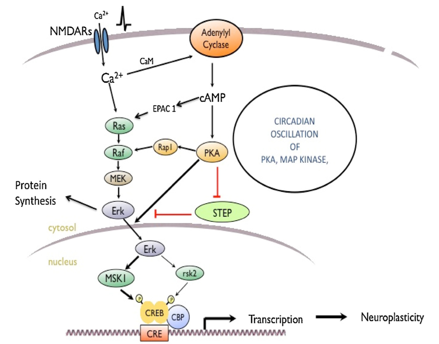
Luckily for glutamate, it’s receptor (NMDAR) is located on the outside of the membrane so it can bind without any issue. Once glutamate binds to NMDAR, NMDAR goes through a change in shape that allows for Calcium ions to travel into the cell. This causes a signaling cascade resulting in the activation of an enzyme called ERK. ERK is then able to bind to the activated GR. This ERK-GR complex then moves into the nucleus (where DNA is stored) and activates two final enzymes MSK1 and Elk-1. These final enzymes cause gene transcription that results in consolidation of memories associated with the stressful trigger.


Application of the cellular mechanism to the real world

Whew! Now that we have the basics understood, we can begin to understand why PTSD occurs. The current hypothesis is that the previously described pathway is sent into overdrive by the stressful event. In short, there is too much activation of GR and NMDAR leading to consolidated memories that are too salient. This explains why something as innocuous as an alarm clock can trigger someone’s symptoms of PTSD.

For example, pretend someone has witnessed a horrible, loud car crash. This causes immense psychological stress, and their brain is bathed in a neurochemical slurry of corticosterone and glutamate. This is going to lead to extreme memory consolidation of the car crash. One of the theories on how memories are stored is called the hierarchical model. You can learn more about it here. Basically, the theory posits that thinking of one attribute of something will prime the memory of it (e.g., thinking of wings, flight, and feathers makes one think of a bird).[3] Say one of the things you associate with the car crash is loud noise. Now when you are primed to think of a loud noise by your alarm clock, this triggers the memory of the car crash causing you to re-live that painful moment.[4]

That makes sense! How about treatment? Is there anything we can do to prevent or treat PTSD?

There are a few things one can do to prevent PTSD. The review paper found that rats who voluntarily exercised had increased neurogenesis and had part of the intracellular cascade of PTSD inhibited.1 Several other studies found that increased cytokines (a marker of stress) in the blood of soldiers before they went off to war led to an increased risk of their developing PTSD.[5] This suggests that finding ways to destress and lower cytokines in the body may be neuroprotective for PTSD.

Closing Remarks

PTSD is a widespread issue, but it’s not an insurmountable problem to overcome. As more people learn the underlying cellular mechanism, the more innovative treatments and preventative measures will be discovered. Until then, exercise and reducing stress in our lives seems to be the best way to prevent PTSD.


Sources

[1] Johannes, M. H. M. eR. (2014). Making memories of stressful events: a journey along epigenetic, gene transcription and signaling pathways. Frontiers in Psychiatry, 5. https://doi.org/10.3389/fpsyt.2014.00005

[2] “What Is Posttraumatic Stress Disorder?” What Is PTSD?, American Psychiatrics Association, 2020, https://www.psychiatry.org/patients-families/ptsd/what-is-ptsd#:~:text=PTSD%20affects%20approximately%203.5%20percent,as%20men%20to%20have%20PTSD.

[3] “Top 3 Models of Semantic Memory: Models: Memory: Psychology.” Psychology Discussion – Discuss Anything About Psychology, 11 Mar. 2017, https://www.psychologydiscussion.net/memory/models/top-3-models-of-semantic-memory-models-memory-psychology/3095#:~:text=Hierarchical%20Network%20Model%20of%20Semantic%20Memory%3A&text=are%20organised%20into%20a%20hierarchy,logically%20related%20and%20hierarchically%20organised.

[4] https://www.google.com/url?sa=i&url=https%3A%2F%2Fcommons.wikimedia.org%2Fwiki%2FFile%3AHierarchical_Model_Mental_Lexicon.png&psig=AOvVaw1CxCYu7sAQA30ZG_rZG3lJ&ust=1634767608200000&source=images&cd=vfe&ved=0CAsQjRxqFwoTCNCYrKi-1_MCFQAAAAAdAAAAABAD

[5] Neylan, T and O’donovan, A (2019). Inflammation and PTSD. PTSD Research Quarterly. 29(4).

Understanding The Epigenetics of Addiction

Understanding The Epigenetics of Addiction


Addiction has been a pervasive problem that has plagued human history since we began recording it. Addiction is an issue we are still grappling with, even in developed nations. In the United States, nearly 21 million people have one or more addictions but only 10% of them receive treatment. [1] This costs the US economy north of $600 billion annually and has resulted in 700,000 deaths from overdose between 1999 and 2017.1 These are abysmal numbers. I will try to shed some light on what some of the causes of addiction are, as well as a brief overview of the neurochemistry of addiction.

Neurochemistry of Addiction

Before we try tackling the chemistry, lets introduce the key brain structure in addiction. Arguably, the most important part of the brain in addiction is the nucleus accumbens (NAc). This is the area of the brain that all drugs of abuse act on in some way.[2] All addictive drugs cause an burst of dopamine (DA) release in the NAc. 4 While this is the structure that will be referenced in this article, it should be known that many more areas of the brain play a role in addiction. A comprehensive review can be found here.[3]

Dopaminergic-system-and-reward-processing-in-brain reproduced from PsyPost.


Now to introduce one of the chemical messengers in addiction: FosB. FosB has been found to be expressed after exposure to nearly all drugs of abuse. 4 After repeated drug exposure, the levels of FosB only increase in the brain. As the levels continue to climb, a person’s sensitivity to natural rewards and drugs increases (as does the likely hood of voluntarily consuming more drugs). 4 FosB causes epigenetic changes in the brain, which increase the likelihood that someone will do drugs. This is one of the causes for the vicious cycle of drug abuse.

Causes of Addiction—Epigenetics

I will not try and explain every cause of addiction in this post for two reasons:

    1. It will be a very long list
    2. We don’t know what all of the causes are

However, I will provide a brief overview so we can better understand the causes of addiction, both social and molecular. A recent literature review finds 50% of variance in addiction is dues to genetic variation, while the other 50% is due to environmental variance[4]  First, lets explore the genetic variation, but to do this we have to understand some basic epigenetics.

One of the most fundamental aspects of epigenetics, is the ability of a histone protein to become acetylated (i.e. stick a acetyl group onto a histone). Normally, the histone protein tightly binds DNA and prevents it’s transcription into mRNA. However, when the histone protein is acetylated, it loosens up and allows the DNA to be transcribed (see the following image for a visual).[5]

Epigenetic Modifications. Reproduced from Wikimedia Commons

In addiction, histone acetylation occurs after acute exposure to stimulants like cocaine or amphetamine (meth). [6] Interestingly, these acetylations occur near promoters of c-fos and fosB in the nucleus accumbens (NAc). Earlier in this article, we mentioned how fosB  is one of the molecular causes of promoting addiction. Now we know where it is coming from and why.

Conclusion

Drug addiction is still a problem in modern society. We have seen that epigenetic changes occur in the brain after exposure to drugs of abuse. This causes mental and physical changes to the person that increases their likelihood of continuing to consume drugs. Hopefully through studying the science behind what causes addiction, we can develop new treatments and improve on prevention strategies.

Sources

[1] https://www.addictioncenter.com/addiction/addiction-statistics/

[2] Scofield, M. D., Heinsbroek, J. A., Gipson, C. D., Kupchik, Y. M., Spencer, S., Smith, A. C., Roberts-Wolfe, D., & Kalivas, P. W. (2016). The Nucleus Accumbens: Mechanisms of Addiction across Drug Classes Reflect the Importance of Glutamate Homeostasis. Pharmacological reviews68(3), 816–871. https://doi.org/10.1124/pr.116.012484

[3] https://www.brainfacts.org/-/media/Brainfacts2/In-the-Lab/Animals-in-Research/Brain-Illustration-Reward.jpg?h=367&iar=0&w=650&hash=F19DD0579346B918C0126E49A1FB7B4D32967584

[4] Reul, J. M. (2014). Making memories of stressful events: A journey along epigenetic, gene transcription, and signaling pathways. Frontiers in Psychiatry, 5. https://doi.org/10.3389/fpsyt.2014.00005

[5] https://www.google.com/url?sa=i&url=https%3A%2F%2Ftheory.labster.com%2Fhistone-acetylation%2F&psig=AOvVaw3ybLsvI2s_wjYfMn32Uz1y&ust=1634161272580000&source=images&cd=vfe&ved=0CAsQjRxqFwoTCPjCv6TrxfMCFQAAAAAdAAAAABAT

[6] Renthal, W., & Nestler, E. J. (2009). Histone acetylation in drug addiction. Seminars in cell & developmental biology20(4), 387–394. https://doi.org/10.1016/j.semcdb.2009.01.005

Concussions and sports safety


What is a concussion?

Concussions are a risk that most contact sports pose to athletes; however, concussions can occur from being in combat and unusual accidents. A concussion can be classified as a traumatic brain injury due to physical impact or force on the skull causing damage to the brain. The total impact/force to the brain can stimulate neuronal dysfunction. Some of the symptoms associated with concussion include: migraines, headaches, impaired cognition, and delayed reaction time.

What happens during a concussion?

The total impact/force to the brain can stimulate neuronal dysfunction. Neuronal dysfunction includes increasing the concentration of sodium and calcium entering the cell, as well as increasing the concentration of potassium exiting the cell causing changes in neuronal signaling cascades and neurotransmission. In essence, the cell cannot maintain a typical state of functioning.

In order to compensate for the ionic imbalance, some additional calcium can be stored in the mitochondria. However, this increases metabolism so much that the production of free-radicals can cause even more damage to the neuron over time. The increased calcium concentration in the neuron can also stimulate neurofilament collapse. Neurofilaments are essential to transmit electrochemical impulses down the axon of a neuron and damage results in the diminished ability to transmit messages. Some examples of this include delayed reaction time and memory impairments, which are essential in sports.

Testing:

Many schools require athletes to conduct an ImPACT test online before sports are played to access normal brain function to create a baseline. Having the baseline is important to determine the extent of the damage, such as decreased visual memory and reaction time after a concussion. The online program can also indicate if someone is trying to hide their symptoms in order to return to sports, being handy for a medical professional to access return protocols. However, even if this tool is used to determine if a concussed athlete is ready to play again is debatable. Some studies demonstrate that BOLD (blood oxygen level dependent) increase after a concussion, improving overall cognition, to skew ImPACT testing results to put an athlete who still needs to rest at risk for a re-injury, resulting in even greater symptoms.

Concussions and sports:

The two sports with the highest practice-related concussion rates are football with 5.0 per 10,000 athletic exposures and cheerleading with 3.6 per 10,000 athletic exposures. Football helmets have gotten significantly better to reduce the amount of impact received by players, meanwhile cheerleading does not provide much in terms of safety equipment to protect the heads of athletes. Approximately 70% of concussions in cheerleading come from stunting, many occur from simple mistakes. Not all sports can completely avoid the risk of concussions, but safety and prevention techniques can always and should be improved.

Treatments for concussions:

Treatments for concussions are typically based on supportive services, such reducing screen time and working with a physical and occupational therapists. New research has indicated that the supplementation of DHA, which is an omega-3 fatty acid, which could decrease the recovery time associated with concussions. DHA has neuroprotective effects to help decrease oxidative stress, decrease inflammation, decrease calcium influx within the cell, and improve the integrity of ion pumps and channels as seen in Figure 1. However, the amount of DHA that needs to be supplemented to people with concussions is debatable, which could be due to prolonged poor nutrition, especially in athletes, or actually needing more DHA in the body after an injury. Therefore, having an adequate amount of DHA mitigate neuronal dysfunction as seen in typical concussions.

Despite so many individuals suffering from concussions every year, treatment options remain limited and very little can be done besides treat the symptoms of concussions.

Figure 1: DHA supplementation on brain injuries.

So, what?

Concussions extend beyond a simple hit to the head. Concussions can decrease cognitive function and reaction time, as well as needing to reduce normal activities (using any screens) and increasing the likelihood of developing neurodegenerative diseases later in life. Concussions can cause major distress in athletes who are students, which can impact their education by not being able to catch up on homework in an online capacity. Currently, treatment options are extremely limited and need more work to get people back to their normal functioning after experiencing a concussion.

Sources:

  1. https://pubmed.ncbi.nlm.nih.gov/25232881/
  2. https://neurofilament.osu.edu/research/neurofilaments/
  3. https://www.upmc.com/services/sports-medicine/services/concussion/baseline-testing
  4. https://www.brmh.net/services/orthopedics/athletic-training/concussions-and-impact-testing/
  5. https://publications.aap.org/pediatrics/article/144/5/e20192180/38225/Concussion-Incidence-and-Trends-in-20-High-School?autologincheck=redirected
  6. https://www.ncbi.nlm.nih.gov/pmc/articles/PMC4201839/
  7. https://www.xiahepublishing.com/m/ArticleFullText.aspx?
  8. https://www.chrichmond.org/services/neurosciences/concussion-and-traumatic-brain-injury/concussions-and-technology

NFL working to reduce concussions.

Overview

The NFL is among the most watched sports league in the United States, it is also among the leading sports in diagnosed concussions. In 2019, the NFL had 224 diagnosed concussions over their 17-week season (Battista et al 2020). The NFL has been working to implement changes in the game and equipment to help decrease the number of head injuries each year.


Improvements in evaluation

The improvements in the protocol made since 2018 include, defining impact seizure and fencing responses as independent signs of potential loss of consciousness, representing “No-Go” criteria, requiring an evaluation for all players demonstrating gross motor instability to determine the cause of the instability, and requiring all players who undergo any concussion evaluation on game day to have a follow-up evaluation conducted the following day by a member of the medical staff (NFL players health and safety et al., 2021).


Rule changes

Rules implemented on kickoffs

Since 2019, the NFL has implemented 3 new rules to help reduce the number of concussions. The blindside block is eliminated, expanding protection of defenseless players. It is now prohibited for a blocker to initiate forcible contact with his head, shoulder or forearm when his path is toward or parallel to his own end line. This is more of a technique change since concussions tend to be a bigger issue in the inexperienced NFL players. Another rule change was it is now a foul for running forward and leaping across the line of scrimmage in an obvious attempt to block a field goal. This just means you cannot get a running start in order to block a kick which results in less impact. Their last change wasn’t as much of a change, but they decided to keep the kickoff changes made in 2018 as there was a 35% decrease in concussions on kickoffs as a result (NFL Football Operations et al., 2021).


Equipment changes

The NFL uses Radio Frequency Identification (RFID) tags inside of the equipment which detects impact. These RFID tags help the league to collect more information than ever before about the duration and direction of head impacts players experience based on their positions, both during practices and games. The NFL then breaks down the concussion causing impacts and collects information on the players position, acceleration, and forces. This data has helped the NFL develop position specific helmets to help reduce the number of concussions suffered (NFL et al., 2021).


Conclusion


The NFL is continuing to collect impact data, implement new rules, and creating new equipment standards to help reduce the number of concussions each season. The goal of the NFL is to ultimately eliminate concussions from football; there is still a long way to go.


Works cited


Battista, J. (2020, January 23). NFL reveals 2019 Injury Data, hopeful rule changes are working. NFL.com. Retrieved November 14, 2021, from https://www.nfl.com/news/nfl-reveals-2019-injury-data-hopeful-rule-changes-are-working-0ap3000001098679.


Health & Safety Rules Changes. NFL Football Operations. (2021, August). Retrieved from https://operations.nfl.com/the-rules/rules-changes/health-safety-rules-changes/.


NFL. (2021, August 20). Concussion protocol & return-to-participation protocol: Overview. NFL.com. Retrieved from https://www.nfl.com/playerhealthandsafety/health-and-wellness/player-care/concussion-protocol-return-to-participation-protocol.


Nfl. (2021, October 14). Built by data: NFL Helmet Innovation. NFL.com. Retrieved from https://www.nfl.com/playerhealthandsafety/equipment-and-innovation/engineering-technology/built-by-data-nfl-helmet-innovation.

Pathophysiology of Symptoms after a Concussion

Migraines

Migraines are likely to occur following a concussion for a number of reasons. Whether the concussion was due to an impact or non-impact injury, there has been damage to the brain. Following a traumatic brain injury, the brain is hypersensitive due to the ionic flux and glutamate release. Immediately after an injury to the brain, there is a large release of neurotransmitters and ions. Glutamate binds to NMDA receptors and an increased amount of it will lead to more depolarization in the neurons. Potassium efflux and calcium influx occur which shifts membrane potential. These changes in physiology cause issues for various other cell functions. Figure 1 describes the different types of headaches that can occur following a concussion. The type of headache someone is experiencing can help physicians determine what areas of the brain have been damaged. 

Figure 1: A few of the different types of headaches someone could experience following a concussion. 


Vulnerability to Further Injury 

The sodium-potassium pump must now work harder in order to restore membrane potentials. The Na+/K+ requires ATP, so the body will divert more energy to the damaged brain areas. An increased amount of ATP triggers an increase in glucose metabolism. This increased glucose metabolism will not last for long; there is often a state of impaired metabolism following hyperglycolysis. 

Cerebral blood flow is often reduced following a concussion. The increased demand for ATP along with the decreased blood flow causes further issues in the brain. Energy reserves in the brain are depleted because of the overall lack of energy, oxygen, and mitochondrial function. Metabolic pathways are altered after a concussion and take time to recover. It is unknown how much time is needed for the brain to fully recover following a TBI, but there is a period of vulnerability after it occurs. The altered state of the brain takes time to recover. It is possible for another injury to happen after a concussion and has the potential to cause even worse damage. Figure 2 shows some common areas that are affected after a TBI and what their primary functions are. 

Figure 2: Areas of the brain often damaged during a TBI and what processes they are responsible for. 


Slowed/Altered Cognition 

Damage to the brain following a concussion can lead to a wide variety of symptoms. During a TBI, there is damage to axons. This can occur on a microscale and cause disruption of action potential transport, disconnection, and microhemorrhages. MRI has been used to measure how white matter changes following a concussion. Researchers are able to gain insight into how much axonal damage has occurred by observing white matter. Studies have shown that decreased or damaged white matter after a TBI can lead to a myriad of symptoms such as depression, PTSD, impaired memory, and behavioral changes. 

The parts of the brain responsible for judgment, problem-solving, and arousal are often damaged during a concussion. These physiological changes help explain why many people experience cognitive alterations. People can have increased fear responses and higher levels of depression following a TBI. This video provides a good explanation of common emotional symptoms after a concussion and how to help someone experiencing these changes. 


The Future Has…. Flags?

Free Cartoon Football Players, Download Free Cartoon Football Players png images, Free ClipArts on Clipart Library

Image 1

Ask yourself for a moment, is tackle football really something the youth should be involved in? I know I probably sound like an old geezer tweeting vigorously about “the younger generation” when I say that, but seriously think about it for a moment. People talk about how the brain doesn’t stop developing until roughly the age of 25. Concussions are bad at any point in ones life, but before 25 when rapid cognitive development is occurring, large concussive impacts along with that development seem to be a recipe for disaster. Sure tackle football can teach great life lessons and foster lifelong relationships, but it can also leave those that play it in an altered mental state for the remainder of their lives. Ok, watch the video directly below and then we’ll talk again on the other side.

Video 1

So… did your opinion change? What is an age that you would feel comfortable allowing your own children to start tackle football, if at all?

Is flag football a safer alternative?

American Flag Football Leauge Ultimate Final in EaDo: July 2018 | 365 Houston

Image 2

Lets take a moment to look at some statistics before you judge flag football too harshly. Tackle football players age 6-14 sustained 15 times more head impacts than their flag football counterparts. Head impacts that were considered to be “hard hitting” occurred 25 times more frequently in tackle football than flag football. Essentially, in tackle football head impacts occurred far more frequently and with far greater force. These factors create more opportunity for concussions to occur in tackle football than in flag football. This inherently makes sense, it is the nature of flag football to mitigate collisions.

Ok, lets continue looking into the differences. Flag football players have a median of 8 head impacts per season while tackle football players experience a staggering 378 head impacts per season. These numbers alone should worry any parent who has a child involved in tackle football, think about that number for a second and consider the last time that you had a head impact let alone 378. I like football a lot, my brother and entire family played it, but I would be hesitant to put my future children in harms way. Even if the 378 head impacts are minor they can still compile on one another to create issues. My brother was a lineman in football, he would usually experience relatively few large hits but he would be hit nearly every play on the line for an entire game. The small hits he took throughout the game were so great in number that he sometimes had concussive like symptoms even though he never took a “big hit.” Look at the hit he delivers to the opponent in this play alone, this is not an anomaly is football, rather it is the norm. For reference he’s the big guy in blue in the middle that levels the orange player. Yes he delivered the hit in this situation to parts of the body other than the head, but there were plenty of times that the hits were in the direction of his head.

Video 2; via me

Youth football players can be as young as 5 years old. Think about that, if these same players get to play college ball they have played 13 years of football already. That means they’ve had 13 years to potentially suffer concussions. If they start at, oh lets say 10, then they have 8 years where they are compiling head impacts. Sure, kids will be kids and they will most likely receive injuries regardless of what we do to protect them, but allowing them to play tackle football is exacerbating the amount of those potential injuries.

The amount of head-to-head contact is directly responsible for the rates of concussions within youth athletes, eliminating that impact would alleviate the risk for concussions. As players get older they exert more force on one another and the subsequent rates of a concussion occurring increases. Because they exert more force on one another upon impact they are able to hurt one another in all new ways, not just via direct head-to-head impact. For example a player gets hit so hard that his head “whiplashes” and he gets a concussion. It is not longer the direct head contact that causes the injury but the force itself. The implementation of flag football as an alternative to highschool JV and Varsity football would help to alleviate the impacts on players brains. Sure, tackle football might be more fun to watch then flag football, that is all subjective and a personal opinion. However, one must ask the following; is risking player safety worth it when there seems to be an alternative. When players have a helmet on they feel invincible, the male football players in my school were completely indifferent to what they were doing to their bodies, in particular their brain. If tackle football removed equipment and still had contact it would essentially be rugby, which also has staggering rates of concussion due to high impact on the head. The implementation of flags allows for the alleviation of detrimental contact while still allowing for the beneficial nature of football (i.e. the life lessons, teamwork, and friendships that a sport creates).  Spearing rule for offense – Deep South Sports

Image 3

Historically speaking, money is football and football is contact. College football makes 4 billion dollars a year on only 65 of its teams, even more if you include the other hundreds of colleges involved in the many leagues. In the NFL, one franchise alone is worth 3.5 billion dollars on average. Tackle football certainly sells, and I’m actually not proposing we stop any of it. In fact I sit down every Sunday to watch the Vikings lose in spectacular fashion while simultaneously wishing I hadn’t. What I am proposing is that perhaps there needs to be some form of intervention that protects players before they join college teams. Players should be able to grasp and understand the negative side of football before their heads are at risk and they lose the capacity to do so. Eliminating years of potential concussion can benefit those that move on to future contact football, allowing them to start at an older age with a cleaner cognitive slate.


“Comparing Head Impacts in Youth Tackle and Flag Football.” Centers for Disease Control and Prevention. Centers for Disease Control and Prevention, May 19, 2021. https://www.cdc.gov/traumaticbraininjury/pubs/youth_football_head_impacts.html.

Jacobson, N. A., Buzas, D., & Morawa, L. G. (2013). Concussions from youth football: results from neiss hospitals over an 11-year time frame, 2002-2012. Orthopaedic Journal of Sports Medicine1(7), 2325967113517860–2325967113517860. https://doi.org/10.1177/2325967113517860

Concussions

What is a concussion?

Concussions are also known as mild traumatic brain injuries (TBI). Concussions are an injury to the brain that is caused by a blow or jolt to the head. They can also be caused by a blow to the body that causes the head to move/ shift quickly which in turn causes the brain to shift quickly inside the skull. Due to this shift, there is a disruption to normal brain functioning. The main point is that during the rapid shifting of the brain, neurons are injured due to them being stretched and can potentially be broken. Concussions are more likely to occur in females, as they have smaller, more breakable nerve fibers. 

What happens to the brain during a concussion (mild TBI)? 

  • Ionic flux 
      • Potassium going out, sodium going in → membrane permeability starts to have
        problems due to the mild TBI 
      • Membrane permeability becomes “leaky”: notice images to the right


  • Glutamate release → NMDA receptor 
      • Excess glutamate release means more glutamate binding to its receptors 


  • ATP usage increases via excess glycolysis 
    • Injuries to the neurons causes chemicals to leak in and out of the cells
    • When the membrane permeability is “leaky” → attempting to reestablish all the proper concentration gradients 
    • The chemical leaks destabilize the neurons away from their typical state → Large amounts of ATP are required in order to reestablish the proper concentrations gradients 
  • Energy crisis 
    • Mitochondria is stressed out and overworked due to dealing with all of the excess calcium so ATP is diminished 
      • ROS increases 
    • Diminished blood flow to this part of the brain due to energy crisis 
  • Proteasome activation
    • Calcium also increases proteasome activation → proteasomes help to break down proteins 
      • When cell in a stressed place, it begins the breakdown of things that should not be broken down to begin with
    • There is cytoskeletal damage 
    • Inflammation 
      • Upregulation of cytokines trying to fix some of the damage 
    • Axonal dysfunction

Symptoms of concussion:

  • Migraine
  • Decreased reaction time 
  • Slow cognition 
  • Memory impairment 
  • vulnerability to repeated injury 
  • Hippocampus and cerebral decrease 
  • Changes in protein degradation 
  • Chronic atrophy

Treatment options: 

  • Relative rest 
    • Physical rest; recommended for two days after concussion
    • Mental rest; recommended for two days after concussion 
  •  Complete rest 
    • It is important to note that complete rest (such as very low stimulation areas; dark room with little to no brain stimulation) is not recommended
  • Avoid physical activity
  • When returning to complete activity 
    • Add activities back into daily life routine gradually 
  • Medications
    • Tylenol is recommended during concussions for any pain relief necessary 
    • Ibuprofen and Advil are not advised as they do increase bleeding 

Sources: 

Giza, C. C., & Hovda, D. A. (2014). The New Neurometabolic Cascade of Concussion. Congress of Neurological Surgeons. 

Mayo Foundation for Medical Education and Research. (2020, February 22). Concussion. Mayo Clinic. Retrieved November 14, 2021, from https://www.mayoclinic.org/diseases-conditions/concussion/diagnosis-treatment/drc-203

Sandel , E. (Ed.). (n.d.). What happens to your brain when you get a concussion? Concussion Alliance. Retrieved November 14, 2021, from  https://www.concussionalliance.org/what-happens-to-your-brain

The ImPACT of Concussions

New Theories About Brain Concussions - PhysioFit Physical Therapy & Wellness

Figure 1: A mapping of general symptoms associated with concussions.

What is a concussion?

Put simply, a concussion is the result of a mild traumatic brain injury (mTBI). This is common when the brain bounces off of the inside of the skull because of a great force that is applied to the head in a short amount of time. Common examples come from collisions in sports like football and hockey as well as head injuries resulting from falling or car crashes. The cellular mechanics of these events are pictured below in Figure 2.

Figure 2: A map of what happens on a cellular level when the brain experiences a TBI.

To explain what is happening in Figure 2, it makes to try to follow a chronological timeline after injury. First, because of the injury, holes in the plasma membrane are going to develop. This leads to an ion flux because the axon is no longer to maintain an ion gradient between the inside and outside of the cell. Because of this, an energy crisis is going to occur for 2 main reasons: ion pumps and mitochondria dysfunction. Ion pumps are going to be overworked trying to maintain an ion gradient (which will be nearly impossible because of the holes in the plasma membrane due to the injury), which is important because these are ATP activated channels. So, a lot of energy is going to be used attempting this process. To further propagate this energy crisis, the cell becomes overwhelmed by calcium ions, which it will then deposit in the mitochondria for storage. If there is too much of this calcium deposition, the mitochondria will stop working. This effectively shuts down ATP production by the mitochondria. Instead, the cell must now use glycolysis to produce ATP, which is much less efficient. Overall these events lead to a stressed out cell.

ImPACT Testing - Physical Therapy Innovations - MA

Figure 3: Logo from the ImPACT testing website.

The importance of knowing recovery time

With neural damage as serious as mentioned above, it is very important for people to know when they have a concussion. Subsequent injury during a healing period from an initial mTBI can cause very serious brain damage and an associated longer recovery time. So what is there to test if someone has a concussion? Well one method is ImPACT testing. ImPACT testing is a way to test spatial memory and cognitive speed through a series of tests. It works because the user takes an initial baseline test when they aren’t experiencing a brain injury and then are tested against that baseline when there is a suspected brain injury. However, this method is subject to flawed results as the user could purposefully “tank” their first score so that they pass when they are experiencing a brain injury. The test itself is also subject to flaws. A user taking the test back to back might experience a very different score because of the nature of the test and the time component associate with it.

Magnetic Resonance Imaging (MRI)

Figure 4: Picture of an MRI commonly used in fMRI and BOLD signaling analysis.

What to use instead?

Because of the flaws of the ImPACT test, I propose focusing on an emerging practice of using functional nuclear magnetic resonance imaging (fMRI) to analyze blood-oxygen level dependent (BOLD) signaling in the brain. BOLD signaling analysis works because deoxyhemoglobin (deoxygenated blood) is paramagnetic, causing it to appear on fMRI scans. So, these BOLD signaling studies can show where deoxyhemoglobin is present in the brain, which can be reasonably associated with oxyhemoglobin (oxygenated blood) and its deposition of oxygen in certain regions of the brain. Areas of the brain undergoing rapid repair (such as those that have experienced a mTBI) will need this oxygen. So, by using BOLD signal studies, we could determine a general recovery time associated with a certain level of mTBI experienced. This method is still in development because the association between deoxyhemoglobin and oxyhemoglobin isn’t as simple as I described above and this method is quite expensive.

The future of concussion recovery

By showing the severity of concussions to brain health, it is clear that more needs to be known about how concussion recovery occurs. Having a simply way to measure the pace of recovery, such as using ImPACT testing or fMRI BOLD signal studies, would greatly help. Perhaps there are other methods being developed like fMRI BOLD signal studies that give a more molecularly detailed description of recovery.

Figure 5: Artstract by Trenton Vogt. This figure depicts (very rudimentarily) how spatial memory can be impacted by a concussion.

How are animal models used for Autism studies

Autism is currently one of the most common neurodegenerative diseases, characterized by impairment in communication, social interaction, repetitive behavior, amongst many others. There is no clear notion of the real cause of the disease, however, various animal models has shown various genetic and environmental factors that contribute the autistic symptoms.

Researchers identified involved genes through understanding disruption of the PI3K/Akt/mTOR pathway and possible factors that may have lead to that.

Animal models are a large component of these findings by using rodents such as mice and rats. A large group of people is still against the idea of using animal models for science, which why this blog will be identifying the importance of using animal models, the ethics involved and the important findings they contributed to the understanding of autism diagnosis and treatment.

What to know about animal models:

basic science research

Poor animal care is not good science. There are established US federal laws that the use of non-human animals in research show the efficacy of new treatments.

Models: genetically modified mouse models for ASD-associated genes, were used to learn different facets of ASD at onset, hereditary, therapy, behavior, diagnosis and pathological level.

Challenges: difficulty to generate a mouse model showing all those characteristics, and the differences between mice and humans’ genome.

Example of Animal model for ASD:

For behavior symptoms:

Requires presence of at least six symptoms: two qualitative measures of social impairment, one communication impairment, one symptom for restricted and repetitive impairment.

Social behavior:

Mouse is placed in a open field with an inanimate object and non-familiar mouse and the tendency and time spent with either is measured. The sniffing and following and physical contact with either is measured by video tracker to assess social deficits in autism.

Genetic mutations:

Tuberosis sclerosis:  caused by mutations in the downstream targets of the P13K/Akt/mTOR pathway in the TSC1 or TSC2 genes. It is characterized by seizures, high intellectual impairment similarly seen in autism.


Spam prevention powered by Akismet