“Things I Wish People Knew* Concussions”

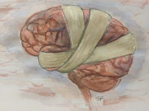
*About (See that’s the concussion doing it’s thing)

For this blog, I will again include potential symptoms as well as neuroscientific aspects behind concussions. However, I first want to focus on some other information provided to me by a friend currently dealing with a concussion. Hopefully this will prove to be equally, if not more, beneficial to you. Wishing her a steady recovery, here are 10 things my friend wishes people knew about concussions:

  1. Every concussion is different. One experience (maybe your own or one that you’ve heard about) is not necessarily generalizable to others’ experiences. It’s really hard to understand what it’s like until you’ve had one.
  2. It may be a moot point, but you can’t see concussions. Therefore, even if someone seems fine, it doesn’t necessarily mean that they are. 
  3. Concussions make everything harder, especially because they never really “turn off” and your head constantly either hurts, has pressure, or feels foggy. While it varies from person to person, concussions can make reading, seeing, writing, listening, thinking, sleeping and, therefore, just existing hard. It makes you feel helpless.
  4. Have compassion, be patient, give endless amounts of love. Having a concussion can make people feel isolated and alone because they have to spend a lot of time in quiet environments, which can often mean by themselves. For people in your life who struggle with mental health, all of that forced alone time can especially take a toll. 
  5. Be there for your loved ones with concussions, but try to avoid frequently asking “How’s your head?” or “How are you feeling?”. Of course, it’s important to show that you care and to let them know that they can talk to you about their struggles any time. But on the other hand, they are constantly dealing with their concussion and probably would appreciate a different topic of conversation once in a while! 
  6. Instead of making people feel guilty, be conscious of people’s concussions because they have no control over the symptoms. Trust that they are fighting and handling life as best they can, even though you may not see it. They are probably already pushing their brain to do more than it should. It’s not their fault that they are not able/ supposed to do certain things (like use technology, watch movies, or be in a loud environment). 
  7. Concussions are not about “just working hard” and “pushing through”. Pushing through is painful, not productive and not helpful in the recovery process. 
  8. Symptoms can rapidly and unexpectedly change. Someone may be feeling alright one minute, but not so much the next. This can be applied to the recovery process as well because it is often not linear. 
  9. Never make the person with the concussion feel like they need to “prove” their injury to you. Believe them. Especially when it has already been diagnosed.
  10. Support them. Love them. Hugs galore (if they like hugs).

After providing this advice based on personal experience, the information below covers some of the medical and scientific aspects of concussions. 

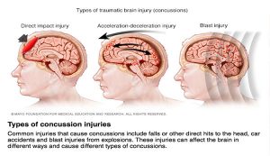
Types, Causes & Symptoms

Causes of concussions are usually significant blows to the head or upper body, sudden acceleration or deceleration of the head, or blast injuries. These also mark the different types of concussions the brain can experience, depending on the nature of the event. Getting any one type of concussion can occur in a wide variety of circumstances.

Image Source

Risks

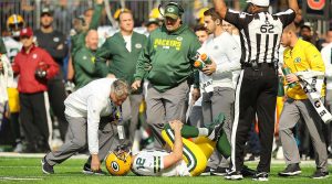
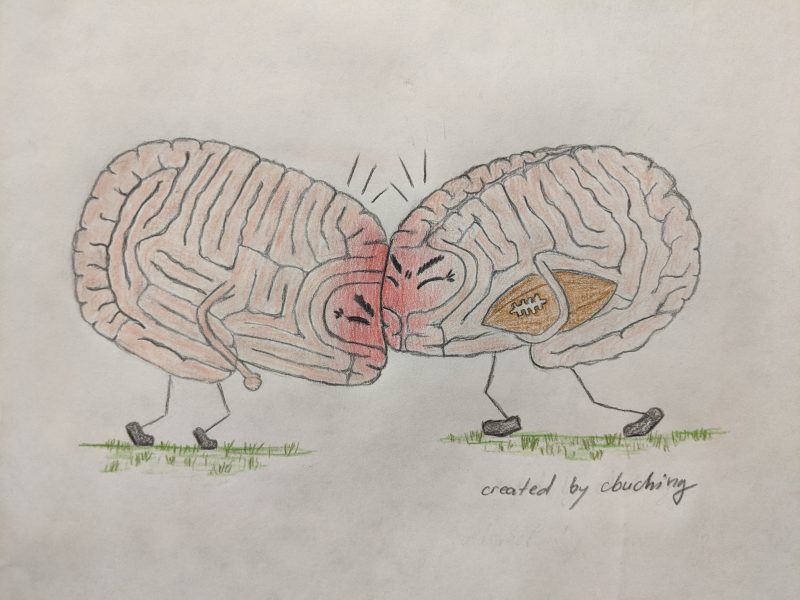
One of the circumstances in which concussions are most well known to occur is football. Lots of research has been conducted for this sport specifically (especially surrounding CTE), but contact sports in general (such as hockey, rugby, soccer, boxing, etc.) pose increased risks for getting a concussion. Furthermore, they are commonly diagnosed in the military as well from events as simple as falling to something as serious as bomb blasts.

Of course the general population can suffer from concussions due to falling as well, but also due to many other incidents like motor vehicle collisions, pedestrian or bicycle accidents, or physical abuse. Having had a concussion before also makes it easier to sustain a second concussion, while possibly making the symptoms more severe as well.

Symptoms

The symptoms and potential consequences of concussions are numerous and may include seizures, loss of consciousness, memory loss, nausea/ vomiting, headaches, mood changes (irritability, anxiety, depression), sensitive vision and hearing, difficulties with physical coordination, memory and concentration issues, lethargic or easily fatigued, trouble sleeping and waking up.

Science of Concussions

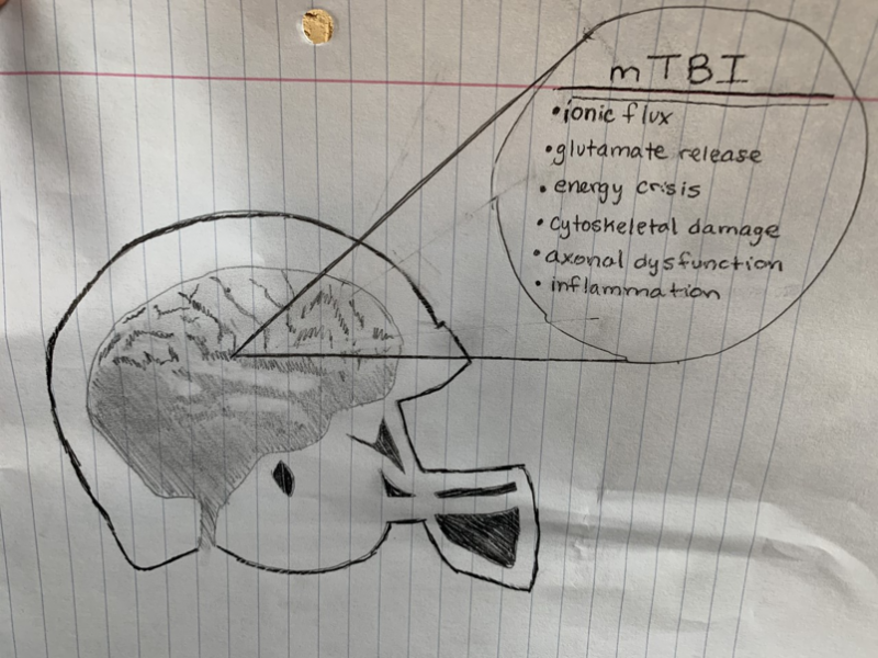
There are many different findings within concussion research as to what the biological/ neural expression of a concussion could look like. One such finding is that mechanoporation (small pores or defects in the membrane) can lead to potassium efflux out of and sodium & calcium influx into the neuron, as well as hyperacute indiscriminate glutamate release. This increased firing of neural cells can then lead to a “depression-like” state, where the firing is less likely, which could be the reason for some of the postconcussive impairments. 

After this increased firing, ionic pumps are actively trying to restore ionic homeostasis of the cell. However, these pumps require energy in the form of ATP (adenosine triphosphate), which means that cellular storage is often strained or even depleted and hyperglycolysis occurs (increased glucose utilization two standard deviations above normal) in order to restore ATP levels. 

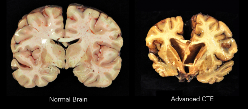
Other pathologies can include cytoskeletal damage, which means that the structural integrity of neurons is lost and in severe cases can lead to axonal disconnection, leading to difficulties in axonal transport. Neurotransmission is also altered due to changes in the composition and function of glutamatergic NMDA receptor subunits. Under some circumstances inflammation and cell death can occur as well. For more information on the science click here.

The SAD American Diet

Obesity rates are skyrocketing in America today and people can point many directions for the cause. Maybe we’ve just gotten lazy? Maybe we don’t have enough time/money to eat healthy? Maybe we aren’t exercising enough? While all these factors are contributing to this epidemic where almost 40% of Americans are obese, there is one biological factor we are missing; how our SAD diet affects our brain.


SAD is an acronym nutritionists have come up with meaning “Standard American Diet”. This diet is characterized by a high intake of processed foods, sugar, fried food, refined grains, high-fat dairy products, and red meat. The Center for Disease Control has found that 9 out of 10 Americans don’t reach the minimum standard for the daily intake of vegetables. Eating all this junk and skipping out on our veggies has led has taken a serious toll on our brains. Not only do physical changes happen in our body, but in our brain too.

After only three days of eating a high-fat diet (our Standard American Diet), scientists found that insulin resistance was reduced in the hypothalamus. Similarly, inflammatory cytokines were inflaming those same pathways. When we develop a resistance to insulin due to inflammation, this leads to an inhibition of PI3K in the insulin pathway. When this occurs, PI3K cannot activate AKT and FOXO1 can’t be removed from the nucleus. FOXO1 in the nucleus inhibits POMC gene expression and inhibits AgRP/NPY. If FOXO is not removed our, AgRP neurons can be stimulated and those are the neurons that make us feel the need to eat. Essentially, when we eat this high fatty diet, we are creating inflammation in our brain that is only encouraging us to eat more and more. It’s a difficult cycle to jump out of.

Many treat obesity as a choice but the science makes it clear there is more at work here than just not having enough will power to stop eating junk food. The food we eat is making changes in our brain, encouraging us to eat more rather than telling us we are satiated.


Studies of obese participants vs non-obese controls have found a number of changes in the brain using neuroimaging techniques. Using fMRI, scientists saw that obese women saw greater activation in their putamen, an important area in the brain for reward when presented with pictures of high-calorie foods and eating utensils compared to lean women. This suggests that obesity has also created heightened responses to visual stimuli regarding food, especially in areas of reward and motivation which is another example of our brain working against us once we are obese to keep us that way.

So, the next time you think obesity is all about choice, make sure you consider the other factors that may be contributing. It’s not as black and white as it may seem. Once you’ve gotten to the point where your brain is actively encouraging you to eat more and responding positively in your putamen to food cues, it’s a tough habit to quit that SAD diet.

https://www.cdc.gov/obesity/data/adult.html

https://www.ncbi.nlm.nih.gov/pmc/articles/PMC3241905/

Obesity Below the Surface

See the source image


Obesity and Inflammation

Inflammation occurs in response to any kind of harmful stimulus. Think of when you get a scratch, the area around it becomes, warm, red, and inflamed. This is a good example of the inflammatory response. However, inflammation can also happen in the brain in response to potentially harmful stimuli. The brain, specifically the hypothalamus can become inflamed in response to overnutrition. This inflammation occurs within a few hours to three days after an overconsumption of glucose or lipids. Inflammation in the hypothalamus can lead to dysregulation of homeostasis. Because inflammation occurs before the onset of metabolic syndrome and related illnesses, it is believed to be a cause of those disorders. https://www.ncbi.nlm.nih.gov/pmc/articles/PMC4389774/#!po=6.19835

Metabolic Syndrome

Metabolic syndrome is a cluster ofSee the source image 
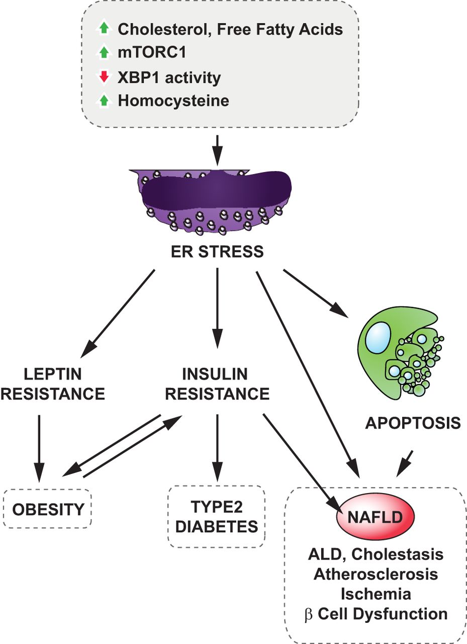
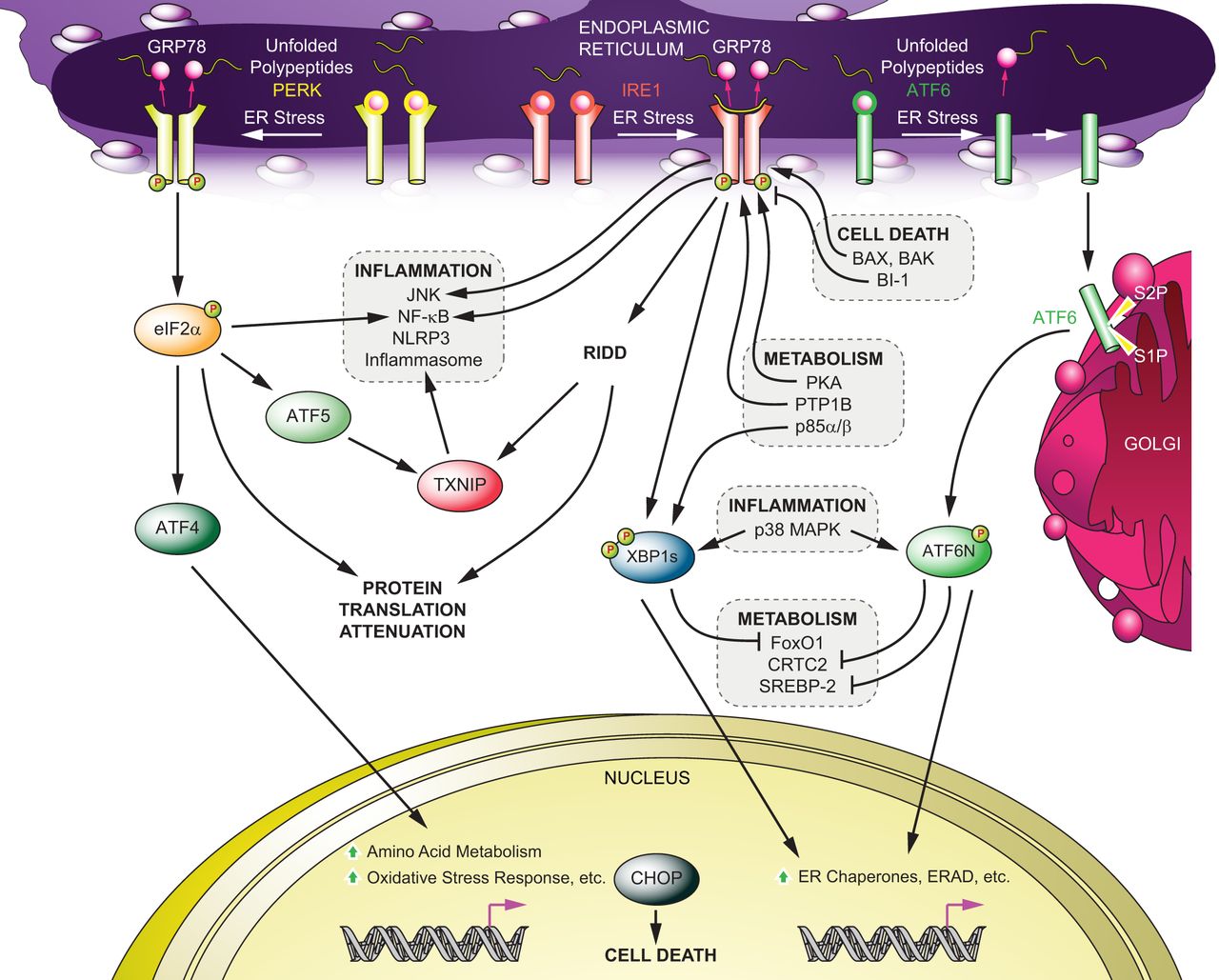
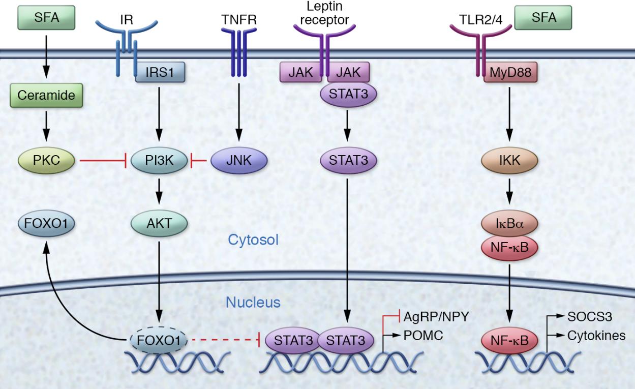
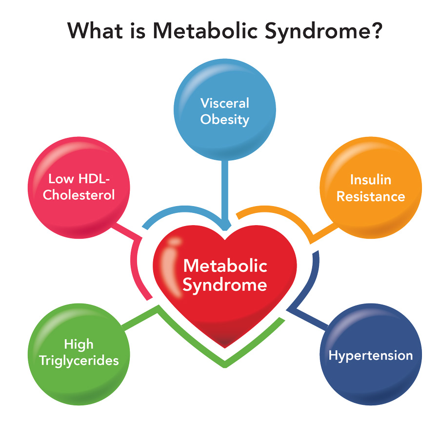
symptoms that lead to an increase in the risk of developing Type 2 Diabetes, Cardiovascular Disease, and stroke. The figure is a good graphic of what components are involved with metabolic syndrome. Having even one component of metabolic syndrome can increase your risk of development of a serious condition. About 1/3 of adults in the United States have metabolic syndrome. One of these components is critical in the hypothalamic inflammation that is seen in obesity, insulin signaling. https://www.mayoclinic.org/diseases-conditions/metabolic-syndrome/symptoms-causes/syc-20351916

Insulin Signaling

An external file that holds a picture, illustration, etc.
Object name is jci-127-88878-g002.jpg

https://www.ncbi.nlm.nih.gov/pubmed/28045396

Insulin resistance is a critical part of obesity and Type 2 Diabetes. When the body becomes resistant to insulin it no longer responds to insulin. The figure above focuses on insulin’s role in the activation of FOXO1. FOXO1 is critical for the activity of POMC neurons (more on that later) and when it is activated it no longer inhibits STAT. FOXO1 can be inhibited by the presence of saturated fatty acids and TNF. The figure also shows another role for saturated fatty acids, downstream activation of cytokines and a protein known as SOCS (suppression of cytokine signaling). This brings us back to the role of overnutrition in the inflammation response. The figure above also shows the role of leptin signaling. In obesity leptin signaling is also dysregulated. Normally leptin is repsonsible for the activation of POMC and the inhibition of AgRP. The last step of this signaling pathway involves STAT which can be inhibited by the inactivation of FOXO1. Obesity also manifests itself in leptin resistance. https://www.ncbi.nlm.nih.gov/pmc/articles/PMC2430504/

POMC and AgRP

An external file that holds a picture, illustration, etc.
Object name is jci-127-88878-g001.jpg

AgRP neuron are regulated through leptin and insulin signaling pathways mentioned above. AgRP neurons are responsible for messages telling us to feed. When this control is lost through leptin resistance, the inhibition is lost and leads to overfeeding. The other half of this control system is the POMC neurons. These neurons are responsible for telling us not to eat. They can be activated by both insulin via FOXO1 and leptin signaling. When resistance to insulin and leptin occurs these neuron become less active. The third part of this diagram shows the importance of the balance between energy expenditure and food intake. When control of POMC and AgRP is loss food intake goes up and subsequently energy expenditure decreases. https://www.ncbi.nlm.nih.gov/pubmed/28045396

There are more pieces than what meets the eye involved in obesity. Once the original overnutrition occurs it triggers changes within the brain. This becomes a vicious cycle of overeating and physiological changes which can be difficult to break out of.


Obesity: Blame the Brain

While diet fads come and go, in the end physicians typically boil there advice down to a simple statement; burn more calories than you eat and you’ll lose weight. Unfortunately, this oversimplification of a very precariously balanced regulatory system in the human body leads to a lot of shame for people who simply can’t keep the weight off. This isn’t a problem facing a few Americans, this is a national epidemic. According to the National Institute of Diabetes and Digestive and Kidney Diseases (NIDDK), 1 in 3 adults are overweight, and 1 in 3 adults are considered obese meaning that a combined 2 out of 3 adults are above their normal weight based on the 2013-2014 survey. This epidemic is far too large to be placing personal blame on every individual. This is not to say that personal choice does not play a role; each and every person must make decisions to eat as healthy as possible within their means as well as dedicating time to exercising. That being said, sometimes its not enough.

What’s the brain got to do with it?

Unlike many common conceptions, obesity is not simply because people are being 
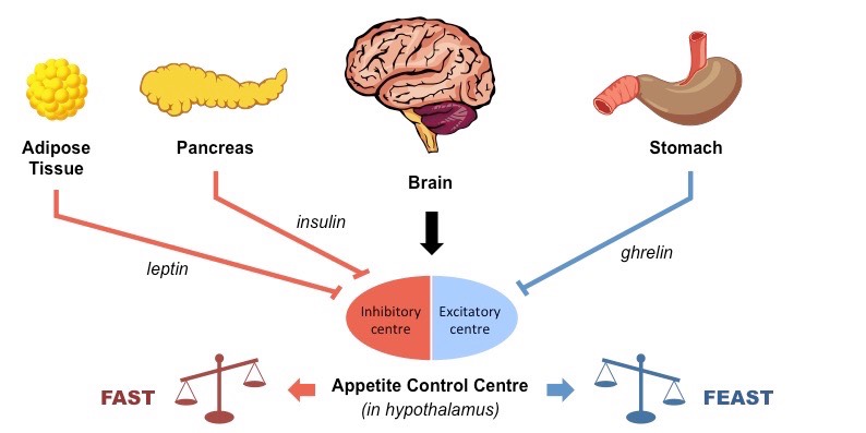
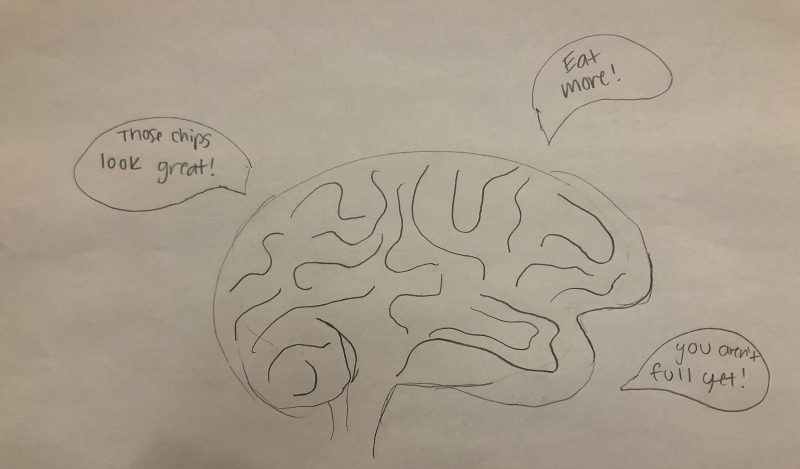
lazy and eating food that is bad for them. While factors such as eating habits, exercise, and screen time all contribute to a person’s weight, so do medications, chronic illnesses, socioeconomic status, and access to healthy foods. Genetic predisposition has also recently been shown to play a major role in a person’s weight. What many people don’t know is that chemical changes in the brain are partially, if not largely, responsible for obesity and can be induced by outside factors.

ER Stress May be Limiting Diet Success

Leptin is one of the main hormones in your body responsible for maintaining balance when you eat.

Lee, J., & Ozcan, U. (2014). Unfolded protein response signaling and metabolic diseases. Journal of Biological Chemistry, 289(3), 1203-1211.

It suppresses appetite and also regulates how much energy your body spends. Working alongside leptin is insulin, which allows your body to use the energy from the food that you eat. When the body becomes resistant to leptin and insulin, energy usage and eating habits get out of whack. New research suggests that endoplasmic reticulum (ER) stress induced by biochemical changes could be responsible. Check out the figure to the right for the data on this. As is shown, when a Western Diet (i.e. high in fat) increases cholesterol and free fatty acids, changes to stress states can result in increased homocysteine and a variety of other changes. This put the ER under stress, which is particularly bad because cells need a functioning ER to maintain proper proteins.

So, what’s the link to leptin and insulin? Well, once the ER is under stress, it sets off a cascade known as the unfolded protein response. In this response shown in the figure, cellular metabolism is altered, and inflammatory actions are dramatically increased.

Lee, J., & Ozcan, U. (2014). Unfolded protein response signaling and metabolic diseases. Journal of Biological Chemistry, 289(3), 1203-1211.

This inflammation, specifically NF-κB proteins, alters DNA transcription and results in the transcription of SOCS, or the suppressor of cytokine signaling. This inhibits specific JAK-STAT signaling, which is the mechanism that the leptin hormone works through. This hormone is tied intrinsically to the insulin pathway, which makes the situation worse. Furthermore, this is just one small problem going on in the cell. Inflammation and other pathways and receptors are being targeted as well, resulting in massive changes to the microenvironment in the hypothalamus, the region of the brain responsible for regulating food intake. These can be seen in the figure below, with the full paper here.

Jais, A., & Brüning, J. C. (2017). Hypothalamic inflammation in obesity and metabolic disease. The Journal of clinical investigation, 127(1), 24-32.


What’s the takeaway?

Obesity is a massively complicated issue stemming from a variety of personal, biologic, and societal issues. It is causing a huge health burden to the world and especially the US. While personal responsibility in making good food and exercise choices is important, exclusive blame can’t be laid at the backs of the patients. Fast food, whether it is in a drive thru line or a box at the grocery store is changing our diets and not for the better. Understanding the complex web of factors that contribute to this crisis is a good first step. Most importantly, changing our narrative about obesity and empowering the medical community to seek out new ideas and solutions is the only way to eradicate this epidemic.


The Brain on a High Fat Diet

Do you ever crave food to the point where you are pretty sure you are addicted? Well, turns out you might actually be addicted to food. Recent studies have connected obesity and overeating to changes that manifest in the brain, specifically linked to energy homeostasis which is the balance of energy in your body. The hypothalamus in your brain is what maintains this homeostasis by regulating feeding behavior and energy expenditure, in other words when you eat and when you exercise is controlled by the hypothalamus. You may be questioning how your brain knows what the rest of your body needs, well this is through insulin and leptin signaling. Insulin and leptin begin signal cascades that lead to the response “stop eating and expend energy.”

Leptin is a hormone produced by adipocytes, fat cells, and binds to receptors in the hypothalamus. The receptor then begins a signal cascade that activates the synthesis of the neuropeptides proopiomelanocortin (POMC) and cocaine and amphetamine regulated transcription (CART). These neuropeptides then lead to the “stop eating and expend energy” response. The leptin signal cascade also results in the inhibition of agouti-related peptide (AgRP) and neuropeptide Y (NPY) which are neuropeptides that signal the “eat now” response. This is great, because it tells your body you’re full. You have enough energy to do life. But there’s a catch! FOXO1 inhibits the action of this signaling cascade leading to the opposite response, “eat more.” To combat this, insulin steps in.

Insulin is a hormone produced in the pancreas, that also binds to receptors in the hypothalamus. Insulin initiates a signal cascade that kicks FOXO1 out of the nucleus so it can’t disrupt the leptin signaling. The figure below shows how leptin and insulin control appetite in the hypothalamus.

Hypothalamus controlling appetite

If there is a lack of leptin or insulin, POMC and CART will not be synthesized to give the “stop eating and expend energy” response. The figure also shows that ghrelin, produced in the stomach will also induce appetite. AgRP and NPY will be synthesized leading to the “eat now” response. If you haven’t eaten for a while and need some energy this is good. But, if this signal occurs even when you’re not hungry, then you have a problem. This uncoupling of the “eat now” response and the amount of energy one has is key in obesity.

So, how does this signal happen when you don’t need energy? Obesity leads to this disruption of energy signaling in a number of ways. One way is that a high fat diet causes the activation of inflammation in the hypothalamus which elicits a stress response from the endoplasmic reticulum (ER). ER stress leads to leptin and insulin resistance, therefore increasing AgRP and NPY neuropeptides and the “eat now” response. The body still produces leptin and insulin, but now the hypothalamus can’t send out the correct signal. This leads to overeating because an individual will continue to eat after attaining their energy threshold. In mice, the hypothalamus returned to its normal size after three days on a low-fat diet. So, no worries, one meal won’t send you down an uncontrollable food tunnel. However, prolonged high fat diets could lead to the uncoupling of energy need and feeding behavior. Therefore, proper nutrition and portions is key to maintaining a healthy lifestyle.

Keep Me in Coach, I’m Ready to Play

In America, it seems that athletics is just as stressed, if not more than, as academics. We live in a culture where professional athletes are praised and put in the spotlight, and the pressure put on them to perform no matter the circumstances is extremely high. So, despite dangerous injuries, professional athletes would often stay in the game to prove their “toughness,” and that tended to be the expectation from coaches since the inception of athletic events. Unfortunately, high school athletes who would follow professional athletes would take cue from them and buy into the idea that they should play at all costs. For specific injuries like concussions, which we now know are extremely dangerous and serious, one important question to ask is why do many high school athletes not report the symptoms of concussion?

Just a Few Reasons

  1. Winning at all costs

High school athletics are important and are most often extremely beneficial to kids with regards to social connections, teaching discipline, and learning vital teamwork skills. However, far too often are high school athletics not kept in perspective.

When asked in a study that was published by the Journal of Athletic Training, up to 55% of high school athletes reported that they didn’t or wouldn’t report a concussion. A strong argument could be made that one contributing factor is the athletes did not want to sacrifice playing time to protect their physical health. They are taken too seriously, specifically when it comes to athletes’ injuries.

    2. “I can’t let my team or coach down”

Although a noble motivation, putting the team or opinion of the coach ahead of one’s physical health is extremely risky. Many athletes feel myriad emotions when it comes to physical injuries, and a common one is that they believe they will look “weak” if they complain of pain. According to an article by Reuters Health, males especially tend to be more sensitive to how their fellow teammates and coaches view them. This is arguably due to the expectation that men are supposed to be strong and resilient, and showing emotion is can be considered a “vulnerability” in the sports realm.

   3. Honest Ignorance

At higher levels of athletics like college and professional sports, the signs and symptoms of concussions are well known and monitored at the slightest instance of a hit to the head.

However, in high school athletics, strict regulations like that of college and professional sports are not uniformly enforced. This leads to high numbers of ill-informed or uninformed high school athletes and coaching staffs. Not knowing the symptoms of a concussion is dangerous, and too many kids have stated that they did not know they had a concussion while they were concussed.

The Solutions

One solution to these problems first and foremost is education. Coaches need to understand the seriousness of concussions and the lasting effects they can have, like difficulty concentrating, personality changes, difficulty with memory, and many more. These are exacerbated if and when someone sustains a second concussion, which is easier to obtain having had an initial concussion. Another point of importance to be stressed to high school athletes, parents of athletes, and coaching staffs is: keep athletics in perspective. Sacrificing enduring brain health problems for a game is never the answer. Brain injuries are unlike injuries to other parts of the body, and injuries to the brain must be treated accordingly. Not to diminish the importance and amazing accomplishments to be had in high school athletics, but after all, it is just a game.

The Science

Many things have been studied that occur after a significant impact to the head is sustained. After impact, potassium flows out of the cell while sodium and calcium flows into the cell, and this is allowed because of defects in lipid membranes that occur after the trauma. This causes an ion flux and a subsequent depolarization that can lead to depression-like responses that are attributed to the post-concussive impairments. To combat this, ionic pumps that require ATP are overactive, which leads to hyperglycolysis. This results in an imbalanced energy supply and demand, which ultimately leads to a variety of metabolic changes that are serious to say the least. The force of an impact can also damage the cytoskeletal structures like dendritic arbors, axons, and astrocytic processes. This often leads to the loss of integrity of axons, and in extreme cases axonal disconnection occurs. Overall, it is not just one specific thing that can be pinpointed when determining the cause for a concussion but rather many things that can and do contribute to the effects of a concussion.

‘Get Your Head in the Game!’ – Figuratively, Not Literally

CONCUSSIONS are one of the most commonly encountered sports injuries where rates are estimated at two million sport related concussions per year in the United States according to the Brain Injury Research Institute. This number is potentially much larger due to the lack of knowledge and awareness about concussion and brain injuries. There has been an increase in research attempting to understand what happens in the concussed brain, but there is a long way to go to help identify treatment plans and return to play protocols. For now, we maybe shouldn’t listen to Zac Efron and his High School Musical buddies. So let’s keep our head out of the game and, instead, focus on raising awareness about concussions and other injuries to the brain.

A vast array of symptoms that can be associated with concussions and traumatic brain injuries (TBI) are a result of the diversity and broadness of factors relating to basic neurobiology and a neurometabolic cascade. These factors include ionic flux and glutamate release, energy crisis, cytoskeletal damage, axonal dysfunction and altered neurotransmission, and inflammation.

IONIC FLUX AND GLUTAMATE RELEASE

Upon a biomechanical injury, potassium efflux and sodium and calcium influx occur due to a damaged phospholipid membrane. An efficient action potential will not be able to be produced because of abnormal depolarization from the rapid influx of sodium and calcium.

ENERGY CRISIS

In an effort to maintain cellular homeostasis regarding ionic flux, ionic pumps that require ATP are shifted into overdrive. This leads to a depletion of energy reserves, thus resulting in reduced cerebral blood flow and an imbalance between energy supply and demand. Prolonged calcium influx leads to increased intracellular calcium and greater amounts of calcium to be sequestered in the mitochondria. Too much sequestration can result in mitochondrial dysfunction which causes the energy crisis.

CYTOSKELETAL DAMAGE

The cytoskeletal structure which include microtubules and microfilaments consist of delicate and complex components. When damage is implemented to a brain region, neurofilaments can easily be phosphorylated and break. Axonal influx of calcium results in the breakdown of proteins into smaller polypeptides on cytoskeletal components. This damage makes the neurons more prone to axonal disconnection and death. After a TBI, ongoing cerebral and hippocampal atrophy may be present. The progressive atrophy can show the appearance of cognitive deficits later in life. The occurrence of a TBI before full recovery from a previous TBI may more likely trigger activation of intracellular proteases and the cascade that leads to apoptotic cell death.

AXONAL DYSFUNCTION AND ALTERED NEUROTRANSMISSION

Increased axonal permeability after a TBI is a result of the vulnerability of the axon. Consequently, this can lead to overall axonal dysfunction. A damaged neuron where neurofilaments and microtubules are altered allows for the neuron to not normally function. Injury to the axon and other white matter areas occurs along with mechanoporation, stretching of individual axons, disruption of axonal transport, axonal misshaping, and axonal disconnection.

Altered neuronal transmission is present after a TBI and occurs due to an imbalance in excitatory and inhibitory neurotransmission. This imbalance is seen due to alterations in the glutamate receptors or GABA receptors which alter excitation and inhibition. Alterations in glutamate, an excitatory neurotransmitter, affect the normal functions of calcium flux and the activation of downstream signal transduction molecules. Decreased levels of GABA, an inhibitory neurotransmitter, affect fear-based learning associated in the amygdala.

INFLAMMATION

Changes in inflammatory markers following a TBI has shown an activation of microglia in the cortex. Microglia function as macrophages in the central nervous system to mediate immune responses. Upon damage to the brain, inflammatory changes are triggered by the upregulation of cytokine and inflammatory genes. Inflammation, although an evolutionary mechanism for protection, has also been shown to induce damage to brain regions that relate to long term side effects. For example, microglial response to damage in the substantia nigra has been shown to increase risk of Parkinson’s Disease after a TBI.

WHY IS THIS SO IMPORTANT?

The research presented above sheds light onto why there are so many symptoms involved with brain injury and concussion. A list of symptoms is listed below:

  • Migraine headache
  • Photophobia and phonophobia
  • Vulnerability to second injury
  • Impaired cognition
  • Slowed processing and slowed reaction time
  • Chronic atrophy
  • Development of persistent impairments

Concussions affect each individual differently and symptoms depend on a number of factors including strength of trauma and the brain area affected by the trauma. When an individual is experiencing these symptoms, they must be monitored closely. Both physical and mental rest can speed the recovery of concussions. Unfortunately, there is still much more research to better understand how to prevent concussions and the symptoms associated with them.

CONCUSSIONS and any injury to the brain are not to be taken lightly. The broadness of symptoms associated can have long term affects. Management of concussions will continue to evolve as more research develops. This will allow us to better understand the steps toward prevention, treatment, and return to play protocols. Protection of athletes and all those more prone to concussion is the number one priority. If you are experiencing any of the concussion symptoms talk to a health professional and play it safe by getting your head out of the game!

For more information on the research presented, follow:

https://www.ncbi.nlm.nih.gov/pmc/articles/PMC4479139/

For more information on the symptoms associated with concussion, follow:

https://www.mayoclinic.org/diseases-conditions/concussion/symptoms-causes/syc-20355594

Why You Should Always Wear You Helmet Even If It Isn’t “Cool”

As a child growing up with a neurologist for a dad, I always heard the phrase “don’t forget to wear your helmet!” whenever I wanted to ride my bike or rollerblade or whenever I took horseback riding lessons. I always thought that it was lame. None of my friends wore helmets so why should I? I always thought they would pick on me or say I wasn’t cool because I had a helmet on. I was embarrassed by it.

Fast forward to now. As a soon to be graduate with a degree in neuroscience, I now completely understand the importance of wearing a helmet whether its simply riding a bike or flying down the slopes on my snowboard.

What happens when you don’t wear a helmet?

Not wearing a helmet increases your risk of brain injury. The most common type of traumatic injuries are concussions. Concussions can occur even with wearing a helmet if the impact is hard enough. Concussions most commonly result from falls, motor vehicle accidents, and sporting accidents.

What happens during a concussion?

Concussions occur when the brain moves inside the skull. After the skull hits a stationary object, that force causes the brain to swirl around inside the skull. The brain bumps into the sides of the skull and damages tissue.

Concussions are not fun. They can cause both temporary and long-lasting problems in cognitive abilities and proper brain function.

Some short-term effects of a concussion include:

  • Temporary loss of consciousness
  • Headache
  • Dizziness
  • Feeling foggy
  • Ringing in ears
  • Nausea or vomiting
  • Blurred vision
  • Sensitivity to light and sound
  • Fatigue

Some long-term effects of a concussion include:

  • Difficulties concentrating
  • Memory issues
  • Irritability or other changes in personality
  • Sleep issues
  • Depression or other psychological problems


Though there is no concussion proof helmet, it is still an important thing to wear. Yes, you can still get a concussion even by wearing a helmet. This happened to me while snowboarding. But wearing a helmet can help minimize the effects. If the helmet can take most of the force, the brain won’t move around as much and not bump into the sides of the skull as hard.


References:

https://healthcare.utah.edu/healthfeed/postings/2016/11/concussion.php

https://www.scientificamerican.com/article/what-happens-to-the-brain/

Image:

https://kidshealth.org/HCA/en/parents/az-concussion.html

Concussion: An Especially Relevant problem in the NFL in 2019

What causes concussion?

Concussions are caused by head trauma, often a blow to the head in which the brain hits the opposite side of the interior of the skull. Common causes of concussion include motor vehicle accidents, falls, and sports injuries. Concussions are classified as mild traumatic brain injury and often characterized by mild cognitive impairments without physical signs identifiable by an MRI or CT scan.

After a concussion occurs, a series of events known as a neurometabolic cascade occurs. The steps occur as follows:

  1. Non-specific depolarization
    1. Cells contain small molecules called ions that normally exist at specific concentrations inside and outside the cell. When these concentrations are disrupted, the charge of the cell changes and causes other events to occur
  2. Release of excitatory neurotransmitters
  3. Potassium efflux
  4. Increased activity of ion pumps to restore the resting state
  5. Hyper glycolysis to generate more ATP
  6. Lactate accumulation
  7. Calcium influx and sequester into mitochondria leading to oxidative metabolism
  8. Decreased ATP production
  9. Overactivity leads to cell death

These changes occur in a cascade one after another and the whole process can have impacts for approximately 7-10 days. These changes make the person especially susceptible to another brain injury. This is where second impact syndrome comes in and why injured people are not supposed to return to their prior activities too quickly. After a concussion, the brain uses all of its resources to repair the damage, so there is nothing else for it to give. When a second concussion occurs, there is nothing left to heal the damage. This is especially important because 50% of all athletes who have experienced second impact syndrome died.

Long term impacts of concussion

A lesser understood risk of concussion is development of a neurodegenerative disorder called CTE. CTE is a form of dementia with similar symptoms to Alzheimer’s disease, but important differences occur. Both disorders can only be diagnosed through autopsy and neither disorder has a cure. Repeated brain injury increases risk for all kinds of degenerative disorders, but each one has its own unique risk factors and these changes are not well understood.

  • Symptoms of CTE include
    • Confusion and agitation
    • Personality changes
    • Erratic behavior
    • Troubles with organization and planning
    • Balance and motor skill problems
  • Symptoms of Alzheimer’s include:
    • Difficulty remembering new information is usually the first sign
    • Gradual increase in level of severity
    • Disorientation
    • Mood and behavior changes (often directed toward family members)
    • Eventually difficulty in life maintaining activities like eating, moving, and breathing

CTE vs Alzheimer’s Disease

CTE Alzheimer’s
Can show symptoms as early as age middle age Most commonly shows symptoms in late adulthood
Tau proteins surround small blood vessels Tau proteins surround B plaques
Catalyzed by repeated brain injury Largely genetic
Lacks diagnostic criteria Relatively well understood symptomology

Especially shocking, 99% of NFL athletes who donated their brains to science were found to have CTE.

To learn more about Alzheimer’s disease, visit the Alzheimer’s Associate website.

Clearly, concussions are dangerous and can have severe impacts. The question then becomes: What do we do about it?

Lately, helmets have been a large topic of discussion surrounding concussions. . From 2016 to 2017, concussions in the NFL increased by 16%. 2017 and 2018 showed a slight decrease in concussions, though the league still recorded well over 200 concussions each year.  As of 2019, the NFL passed new regulations that required all players to wear an approved helmet to engage in any kind of practice or games on the field.  The regulation was the first of its kind as players could “grandfather in” helmet types that had been used in the past. Before this, helmets had not changed significantly since the 70s.

Current relevance

On November 15th, 2019 Myles Garrett hit Mason Rudolph with his own helmet during the final seconds of the third quarter in a game between the Steelers and the Brown. The whole story as well as video is available here.  After hearing some of the science behind head injury, you may see this story a little differently. Are there viable changes that can be made to the sport or is head injury simply and inevitable consequence of playing professional football?

Too Much Protection?

The increasingly competitive nature of sports is constantly pushing athletes to the limit of human capabilities. This provides a more enjoyable sporting event for the fans, but has the potential to have several adverse effects on the athletes. Major league sports has seen a dramatic rise in many types of injures such as torn ACL’s and Tommy John replacement because of the physical demand now being placed on individuals. In recent years however there has been a rise of another kind of injury, one which often goes undiagnosed until after the athlete has completed his or her career. This disease is CTE and is caused by repeated trauma to the head. These repeated concussions lead to changes of behavior, cognitive function, physiologic function. In order to better understand concussions, many researchers are now examining the neurochemistry of the disorder in order to find potential cures and remedies for CTE.

Much of the neurochemistry of concussions is still unknown and research is constantly being done to better understand what is happening within the brain. Some things are known however. Upon the initial the membranes of neurons can become damaged. This leads to calcium ions to enter the cells in concentrations much higher than normal leading to a depolarization as potassium leaves the cell. It is hypothesized that these ion fluxes could be one of the main causes of migraine in regards to concussions. Also, the mitochondria of these synaptic nerves then absorbs this calcium which leads to an energy crisis within the cell as these organelles attempt to restore homeostasis. This lack of energy within the cell can lead to vulnerability of the individual to receive concussions more easily. Energy depletion and abundant calcium within the cell then leads to the alteration of normal neurotransmitters, changing the wiring within the brain. Repeated concussions lead to permanent rewiring of the brain thus leading to the changes already discussed with CTE.

This more complete understanding of concussions has allowed for the advancement of safety equipment in many major league sports. The NFL is a prime example of such innovation when it comes to safety equipment and concussion protocol. Every year the NFL makes alterations in helmet design and regulation, in an attempt to protect their players. However, there are some individuals who do not support all of the advances in safety regulation. Many of these people make the argument that these safety regulations are changing the nature of the sport. For example boxing is a sport, MMA, and UFC are all sports which thrive off of combat. However, is it right to make individuals use more protective gear when the nature of the sport is obviously human-human combat? Also, the individuals partaking in the sport have a voice? If an individual knows the risk of a sport and deems the risks worth the reward who has the right to stop them from potentially being harmed.

The world of sports is rapidly changing. These changes may be more interesting for the audience but can take a demanding toll on an athletes body. With this rise in completion there is also a rise in injury. Knowledge of concussions has been essential for the advancement of safety equipment utilized to keep athletes safe, but how much is too much? Providing to much safety equipment can change the nature of a sport which has a variety of implications. If athletes are aware of the issues shouldn’t they be allowed to take the risks? Finding the balance between safety to prevent concussions while still being true to the sport will be no easy task.

http://www.nfl.com/news/story/0ap3000000897109/article/nfl-implements-modifications-to-leagues-concussion-protocol

https://www.google.com/url?sa=i&source=images&cd=&ved=2ahUKEwi8hsCXgvflAhUC2aQKHS4OCPsQjRx6BAgBEAQ&url=https%3A%2F%2Fwww.semanticscholar.org%2Fpaper%2FNeurochemical-cascade-of-concussion.-MacFarlane-Glenn%2Ff6dac7da9880f4342149fca509152a721ebac3da%2Ffigure%2F1&psig=AOvVaw27dkmz66iRtlDxNmmj9E0Q&ust=1574278264854460

https://moodle.cord.edu/pluginfile.php/798940/mod_resource/content/3/2014%20The_New_Neurometabolic_Cascade_of_Concussion.3.pdf


Spam prevention powered by Akismet