It’s about time you’ve met your metabolism

The article we have covered in a previous week, “Hypothalmic inflammation in obesity and metabolic disease” by Alexander Jais and Jens C. Brüning was an article about metabolic inflammation being connected to obesity in many surprising ways. Basically, we know already that obesity is an obstacle for the brain and it’s functioning. However, due to this feature, this is in a way how this correlation works.The topic today is why people should care about this topic and what the people must know, so without further ado let’s get reading!

The article informs us of a typical metabolic homeostasis, and then what happens to it under insulin and leptin resistance. Though, the article specifically focuses mainly on the insulin and leptin resistance aspect. As a result of this connection, we can now at least suspect the connection!

Figure one, pictured above, from the article mentioned above is especially excellent at explaining this where it was needed (excellently timed, or in other words placed well). That piece is a diagram expressing exactly this. The myriad, but consistent colors complements the black parts which either crosses something out, or is a metaphorical old-timey scale. Although, one slight problem could be that it could feel a large bunch overwhelming if you’re not too familiar with the applied terms of figure one in the Neuroscience field. This was an excellent piece to me for it is maximized simplicity because, for clear reasons, that kind of thing strongly helps. The figure may also benefit people uninvolved in Neuroscience as well because figure one uses so much brief, yet descriptive, labeling, and I find that effective myself in general because it’s easy on the eyes to track or logicate.

Now, at this point, one, such as yourself, may wonder why people really should care about all the above information. Well, let’s answer with essential basics to answer ourselves by quickly asking ourselves something simpler first; what really is metabolism? Well, the answer is absolutely nothing short of deeply important. According to Arturo Sánchez López de Nava and Avais Raja, metabolism is known as “the whole sum of reactions that occur throughout the body within each cell and that provide the body with energy.” (Arturo Sánchez López de Nava and Avais Raja 2022).^2 Considering what we know about energy, the ability to do action internally and externally, it’s no shock to pretty much anyone that such a scenario can turn serious fast. Let us put this into perspective with another topic.

In my class, I personally examined the various effects that the person with a penis has in the impact of their child’s birth via their diet. Surprisingly, I learned from the University of Tennessee Institute of Agriculture, even very specific nutritional choices have a direct effect on the child. Using protein as an example, a low amount of protein may cause a higher birth weight while high amounts of protein may lead to glucose intolerance in their child.^3 It is little short of incredible how essential our nutritional choices are, and even more so that we seemingly take little worry to what it does to us.

References:
1) “Hypothalmic inflammation in obesity and metabolic disease” by Alexander Jais and Jens C. Brüning
2)https://www.ncbi.nlm.nih.gov/books/NBK546690
3) https://animalscience.tennessee.edu/wp-content/uploads/sites/7/2022/05/Relevant-Repro-Blog-7_-Paternal-Impacts.pdf

Rewire Your Metabolism: How Healthy Fats Heal Insulin and Leptin Resistance

Figure 1: Illustrates how brain inflammation contributes to leptin and insulin resistance, with unsaturated fats helping to reduce inflammation and improve metabolism. Created by Sharleen Mtesa.

We’ve all heard the saying “you are what you eat,” but when it comes to hormones like insulin and leptin, that couldn’t be more true. These two messengers have a big job: insulin helps manage your blood sugar, and leptin tells your brain when you’re full. These two hormones play a critical role in keeping our metabolism on track. And when they’re working properly, they help us maintain a healthy balance between hunger and energy.

But when our diets are high in processed foods, unhealthy fats, and sugar, these hormones can stop functioning correctly. That’s when insulin and leptin resistance kicks in leading to constant cravings, weight gain, and blood sugar issues.

Therefore, incorporating healthy unsaturated fats into your diet like those found in olive oil, avocados, nuts, and fatty fish can be a game-changer. These fats help reduce inflammation, improve hormone sensitivity, and support your body’s ability to manage hunger and energy more effectively. Let’s take a closer look at how these fats actually work to restore balance and boost your health.

How Inflammation Disrupts Insulin and Leptin Resistance.

According to the paper “Hypothalamic Inflammation in Obesity and Metabolic Disease,” a high-fat diet (HFD) triggers inflammation in the hypothalamus, impairing how the brain responds to key hormones like insulin and leptin. This inflammation leads to insulin resistance, which makes it harder for the body to manage blood sugar, and leptin resistance, which disrupts the brain’s ability to signal when you’re full, ultimately increasing hunger and food intake.[1]

The inflammatory process involves the activation of proinflammatory cytokines, like TNF-α, that interfere with insulin and leptin signaling. This damage to hormone function results in metabolic imbalances, weight gain, and constant cravings. These effects on hunger and energy regulation create a vicious cycle, making it harder to maintain a healthy weight and manage blood sugar levels

How Unsaturated Fatty Acids Help Reverse Insulin and Leptin Resistance.

Unsaturated fatty acids, particularly monounsaturated fats (MUFAs) and polyunsaturated fats (PUFAs), play a very important role in restoring metabolic balance. These healthy fats not only help with weight control but also improve the body’s response to insulin and leptin by reducing inflammation and promoting fat metabolism.

  • Monounsaturated fats (MUFAs): Found in olive oil, nuts, and avocados.
  • Polyunsaturated fats (PUFAs): Especially omega-3s and omega-6s, found in fatty fish, flaxseeds, and walnuts.

1. Improving Insulin Sensitivity.

Insulin helps cells absorb glucose from the blood, but when the body becomes resistant, blood sugar levels rise, contributing to type 2 diabetes. Unsaturated fats aid in restoring insulin sensitivity in the following ways:

  • Better Cell Membrane Function: Unsaturated fats become part of cell membranes, making them more flexible and fluid. This improves insulin receptor function, allowing glucose to enter cells more efficiently.
  • Reduced Inflammation: Chronic inflammation, often caused by a diet high in saturated fats and processed foods, disrupts insulin signaling. Omega-3s have powerful anti-inflammatory effects, reducing inflammation and improving insulin function. These omega-3s help calm the immune system, prevent activation of inflammatory receptors, and reduce the production of TNF-α, a cytokine that contributes to insulin resistance.
  • Fewer Toxic Fats in Organs: Excess fat storage in organs like the liver and muscles leads to insulin resistance (lipotoxicity). Unsaturated fats help reduce fat accumulation and promote healthy fat metabolism, encouraging the body to use fat for energy instead of storing it in organs. For example, oleic acid (found in olive oil) and omega-3s help reduce diacylglycerol (DAG) buildup in the liver, which is linked to insulin resistance.
  • Gene Regulation: Unsaturated fats activate proteins (like PPARs) [2] that regulate how the body uses sugar and fat, improving glucose control and reducing fat storage.

Figure 2: Insulin Signaling Pathway
This figure shows how insulin activates signaling pathways like PI3K/Akt and MAPK to promote glucose uptake, glycogen synthesis, and cell growth, while SOCS proteins regulate the response through negative feedback.[3]

2. Restoring Leptin Sensitivity

Leptin is produced by fat cells and signals the brain when you’re full. However, in leptin resistance, the brain doesn’t respond, leading to overeating and fat storage. Unsaturated fats help restore leptin sensitivity by:

  • Calming Brain Inflammation: High-fat, high-sugar diets cause inflammation in the brain, especially in the hypothalamus, where leptin acts. Omega-3s help reduce this inflammation, allowing leptin signals to reach the brain effectively.
  • Helping Leptin Cross the Blood-Brain Barrier: Leptin travels from the blood to the brain across the blood-brain barrier. Chronic inflammation can disrupt this process. Omega-3s help improve blood-brain barrier function, enabling leptin to reach the brain more efficiently.
  • Improving Leptin Signaling: Unsaturated fats support better leptin receptor function and reduce the expression of SOCS3, a protein that blocks leptin signaling. As a result, the brain can detect leptin properly, helping to reduce hunger and cravings.[4]

Figure 3: Overview of Leptin Signaling Pathway.
This figure shows the leptin signaling pathway, where leptin activates JAK2, leading to phosphorylation of key sites (pY985, pY1138) and triggering downstream signals like STAT3, PI3K, and ERK. These pathways regulate hunger and energy balance, with SOCS3 acting as a feedback inhibitor.[5]

Feed Your Hormones Right.

When it comes to balancing your metabolism, hormones like insulin and leptin are key players and what you eat has a huge impact on how well they work. Chronic inflammation from highly-processed foods and unhealthy fats can throw these systems off, leading to cravings, weight gain, and blood sugar spikes.

But the good news? Healthy unsaturated fats like those found in olive oil, avocados, nuts, and fatty fish can help reset the system. By calming inflammation, improving hormone signaling, and supporting better fat metabolism, these fats make it easier for your body to respond to hunger and energy cues the way it’s supposed to.

So instead of fearing fat, focus on the right kinds. A few smart swaps in your daily meals can go a long way in supporting hormone health, boosting energy, and helping you feel your best from the inside out.


Footnotes

[1] Jais, A., & Brüning, J. C. (2017). Hypothalamic inflammation in obesity and metabolic disease. The Journal of clinical investigation, 127(1), 24–32. https://doi.org/10.1172/JCI88878

[2] Varga, T., Czimmerer, Z., & Nagy, L. (2011). PPARs are a unique set of fatty acid regulated transcription factors controlling both lipid metabolism and inflammation. Biochimica et biophysica acta, 1812(8), 1007–1022. https://doi.org/10.1016/j.bbadis.2011.02.014

[3],[5] Howard, J. K., & Flier, J. S. (2006). Attenuation of leptin and insulin signaling by SOCS proteins. Trends in Endocrinology & Metabolism, 17(9), 365–371. https://doi.org/10.1016/j.tem.2006.09.007

[4] Park, H. K., & Ahima, R. S. (2014). Leptin signaling. F1000prime reports, 6, 73. https://doi.org/10.12703/P6-73


Your Brain on Fat: Why Hypothalamic Inflammation Matters

Most of us know that a high-fat, high-sugar diet can lead to weight gain. Many people are aware of how obesity is linked to type 2 diabetes, heart disease, and other chronic conditions. But here’s something less commonly known: the food we eat can trigger inflammation in the brain within just a few days, before we even see a change on the scale.

That’s the core insight from the article “Hypothalamic inflammation in obesity and metabolic disease”. This research shows that the brain, particularly the hypothalamus, isn’t just passively responding to weight gain. Instead, it plays an active role in the development of obesity and related diseases.

The Hypothalamus: A Key Player in Energy Balance

Fig 1
Fig 1 [1]

The hypothalamus is a critical region of the brain responsible for regulating hunger, satiety, and overall energy expenditure[1]. As shown in Figure 1, it contains two key types of neurons: POMC neurons, which help reduce appetite, and AgRP neurons, which stimulate it. Under normal conditions, these neurons respond to signals from hormones like insulin and leptin to maintain balance between energy intake and use[1].

However, diets high in saturated fats—particularly palmitic and stearic acids—can disrupt this system quickly. These fatty acids can cross the blood-brain barrier and accumulate in the hypothalamus. Once there, they activate inflammatory pathways like TLR4 and NF-kB, triggering cellular stress and impairing the brain’s ability to respond to leptin and insulin[1]. Essentially, the brain stops receiving the “I’m full” signal, which promotes overeating and weight gain, even before visible changes in body weight occur[1].

Type 2 Diabetes

Type 2 diabetes occurs when the body doesn’t use insulin properly, causing blood sugar levels to rise [2]. This is due to the pancreas producing too little insulin and the body’s cells not responding well to it. Over time, high blood sugar can damage the eyes, kidneys, nerves, and heart[2].

Although once known as “adult-onset diabetes,” type 2 can now affect children, largely due to rising obesity rates[2]. While there’s no cure, the condition can often be managed with weight loss, healthy eating, exercise, and, when needed, medications or insulin therapy[2].

Common symptoms may develop gradually and include:

  • Increased thirst and urination

  • Excessive hunger

  • Weight loss

  • Fatigue

  • Blurred vision

  • Slow-healing sores

  • Frequent infections

  • Tingling or numbness in hands/feet

  • Darkened skin patches, especially in the neck or armpits

Leptin, Obesity, and Type 2 Diabetes

Leptin, a hormone produced by fat cells, plays a central role in appetite control, energy balance, and glucose regulation[3]. In a healthy system, leptin levels rise as fat stores increase, signaling the brain to reduce food intake and increase energy expenditure[3].

But in obesity, something goes wrong: leptin levels are high, but the brain becomes resistant to its effects. This leptin resistance means the brain doesn’t register fullness, which contributes to increased food intake and, over time, insulin resistance a key feature of type 2 diabetes (T2DM). Leptin acts through several neuronal pathways, including GABAergic neurons and cells in the ventral premammillary nucleus of the hypothalamus, though the exact locations and mechanisms are still being studied [3].

Why This Matters

This research challenges the idea that obesity is simply the result of overeating or a lack of willpower. Instead, it highlights a deeper issue: neuroinflammation. The food we eat doesn’t just affect our waistline—it changes the way our brain functions. Inflammation in the hypothalamus can alter how we process hunger and fullness, pushing us toward patterns of overeating and disrupted metabolism.

Understanding this connection opens the door to new therapeutic strategies. If we can find ways to reduce brain inflammation or restore leptin sensitivity, we might be able to intervene early in the development of obesity and metabolic disease, not just treat the symptoms after the fact.

[1]Jais A, Brüning JC. Hypothalamic inflammation in obesity and metabolic disease. J Clin Invest. 2017 Jan 3;127(1):24-32. doi: 10.1172/JCI88878. Epub 2017 Jan 3. PMID: 28045396; PMCID: PMC5199695.

[2] Mayo Clinic. (2025, February 27). Type 2 diabetes. Mayo Clinic. https://www.mayoclinic.org/diseases-conditions/type-2-diabetes/symptoms-causes/syc-20351193

[3]Manglani, K., Anika, N. N., Patel, D., Jhaveri, S., Avanthika, C., Sudan, S., Alimohamed, Z., & Tiwari, K. (2024). Correlation of Leptin in Patients With Type 2 Diabetes Mellitus. Cureus, 16(4), e57667. https://doi.org/10.7759/cureus.57667

Pattern Separation in the Dentate Gyrus

Figure 1. Impact of stress and anxiety on pattern separation.
Created by Sharleen Mtesa. The image shows a brain with disrupted signals and emotional shadows, symbolizing how stress affects memory. Puzzle pieces and swirls represent confusion, while the background figure reflects emotional burden.

Have you ever noticed how certain memories, especially those tied to fear, stress, or strong emotions, seem impossible to forget? These moments, whether terrifying, traumatic, or deeply moving, often stay with us for life. Scientists have found that a small part of the brain called the dentate gyrus (DG) plays a major role in this [1]. The DG, located in the hippocampus, processes incoming information and helps determine which memories are stored long-term, particularly those linked to strong emotions. 

But memory isn’t just about storing emotional experiences, it’s also about keeping them distinct. When stressful or emotional events are similar, how does the brain prevent confusion between them?

Therefore, one key function of the DG is pattern separation, the brain’s ability to transform similar experiences into distinct, non-overlapping memories.

Figure 2. The location of the dentate gyrus in the brain.
This image shows where the dentate gyrus is located inside the hippocampus, a part of the brain involved in memory and emotion.[2]

How the Brain Keeps Stressful Memories Separate

According to the paper “Making memories of stressful events: a journey along epigenetic, gene transcription, and signaling pathways”, the DG achieves pattern separation through a very selective process. Only a small number of neurons in the DG become active during a stressful experience. This phenomenon, called sparse activation[1], helps keep memories sharp and distinct.

But this selectivity isn’t random, it’s carefully controlled by molecular switches within the brain cells. One of the most important switches is a histone tag called H3S10p-K14ac. Think of it like a key that unlocks tightly packed DNA, allowing important memory-related genes, like c-Fos and Egr-1, to be turned on. Without this tag, these genes remain inactive.[1]

Even though stress activates many parts of the brain, only DG neurons with this specific histone tag begin recording the memory. This ensures that the brain focuses its response on just a few key cells, helping to preserve the distinctiveness of each memory.

How the Dentate Gyrus Works Its Memory Magic.

The DG has some cool tricks to keep your memories sharp and organized:

1. Sparse Coding
  • Only a small group of neurons fire for each experience.
  • ➡️ Prevents similar memories from overlapping.
  • Example: Meeting two people with similar voices? DG helps you tell them apart.
2. Input from the Entorhinal Cortex (EC)
  • EC sends sensory + spatial data to the DG.
  • DG reshapes it and passes it to the hippocampus.
  • Ensures detailed, clear memory storage.
3. Inhibitory Microcircuits
  • GABAergic interneurons = your brain’s memory traffic cops.
  • Prevents too many neurons from firing at once.
  • Example: Helps you remember where you actually parked today, not yesterday.
4. Neurogenesis = Fresh Memory Power
  • DG keeps growing new neurons, even in adulthood.
  • These newbies help make fresh, flexible, and distinct memories.
  • Keeps your brain adaptable, not stuck in the past.

Figure 3. Simplified Flow of Information in the Hippocampal Formation.
This figure shows how sensory input from cortical association areas is processed by the parahippocampal and perirhinal cortices, then transmitted to the entorhinal cortex, which relays it to the hippocampus. Within the hippocampus, the dentate gyrus (DG) acts as the initial processing stage, playing a key role in pattern separation, transforming similar inputs into distinct memory representations before passing information to CA3, CA1, and the subiculum. [3]

How Stress & Anxiety Mess With the DG’s Memory Magic.

Stress doesn’t just make you feel awful, it actually affects how your brain remembers.

  1. Stress Hurts Neurogenesis
  • DG produces new brain cells throughout life (neurogenesis).
  • Chronic stress = high cortisol (a hormone the body releases under stress) = fewer new neurons. [4]
  • Fewer neurons → DG can’t keep memories separate → confusion & memory blending.
     2. HPA Axis Overdrive Weakens DG
  • When stress continues for too long, the HPA axis(the body’s stress response system) stays overactive, keeping stress hormones high. [5]
  • High cortisol levels weaken connections between neurons in the DG.
  • ➡️ This makes it harder for the brain to separate new experiences from past ones
  • Leads to anxiety, fear, and biased memory recall.
  1. Pattern Completion Takes Over
  • DG usually favors pattern separation.
  • Under stress, it shifts to pattern completion (retrieving old memories based on partial cues).
  • What that looks like:
    • Fear Overgeneralization: Mistaking safe situations for dangerous ones.
    • Exaggerated Emotional Reactions: Past trauma colors present experiences.
  1. Memory Interference: Past vs. Present
  • DG normally prevents interference between old and new memories.
  • Stress disrupts that filter:
    • Past memories bleed into new ones.
    • Makes it hard to feel safe even when you are. [6]

How to Boost DG Function & Beat Stress

Want to keep your DG happy and your memories sharp? Try these:

  • Exercise: Boosts neurogenesis + lowers cortisol
  • Mindfulness & Meditation: Calms your stress response system
  • Cognitive Behavioral Therapy (CBT): Helps reframe overgeneralized fears
  • SSRIs/Antidepressants: Improve memory-related brain plasticity

Final Thoughts: Protecting Your Brain’s Memory Filter

The dentate gyrus may be small, but its role in shaping how we process and separate emotional memories is huge. When working well, it helps us distinguish the past from the present, keeping our memories organized and our reactions in check.

But chronic stress and anxiety can disrupt this process, blurring lines between memories, fueling fear, and making it harder to feel safe in the moment. The good news? The DG is flexible and responsive. With habits like exercise, mindfulness, therapy, and proper treatment, we can support brain health and improve memory clarity.

Because memory isn’t just about holding on, it’s about understanding, adapting, and moving forward. 


Footnotes:

[1] Reul J. M. (2014). Making memories of stressful events: a journey along epigenetic, gene transcription, and signaling pathways. Frontiers in psychiatry, 5, 5. https://doi.org/10.3389/fpsyt.2014.00005

[2] Centre of Excellence for Early Childhood Development. (2008, December 5). Glossary – Brain. Encyclopedia on Early Childhood Development. https://www.child-encyclopedia.com/sites/default/files/docs/glossaire/Glossary_Brain_DG.pdf

[3] Weilbächer, R., & Gluth, S. (2016). The interplay of hippocampus and ventromedial prefrontal cortex in memory-based decision making. Brain Sciences, 7(1), 4. https://doi.org/10.3390/brainsci7010004 

[4] Schoenfeld, T. J., & Gould, E. (2012). Stress, stress hormones, and adult neurogenesis. Experimental neurology, 233(1), 12–21. https://doi.org/10.1016/j.expneurol.2011.01.008

[5] Lee, J. W., & Jung, M. W. (2017). Separation or binding? Role of the dentate gyrus in hippocampal mnemonic processing. Neuroscience & Biobehavioral Reviews, 75, 183-194. https://doi.org/10.1016/j.neubiorev.2017.01.049 

[6] Kim, E. J., Pellman, B., & Kim, J. J. (2015). Stress effects on the hippocampus: a critical review. Learning & memory (Cold Spring Harbor, N.Y.), 22(9), 411–416. https://doi.org/10.1101/lm.037291.114 


Protecting Your Brain to Protect Your Body: A New Angle on Preventing Obesity

Abstract created by Gabe Sparks

Obesity is often labeled as a simple imbalance between the calories we consume and the calories we burn, but this disease runs much deeper than public perception suggests. It’s not just about food intake; it’s also about complex processes happening within the brain. Recent research shows that inflammation in certain brain regions can disrupt normal signaling in neural pathways, contributing to the development of obesity.1

One of the most affected areas is the hypothalamus, a region that plays a critical role in regulating feeding behavior and energy expenditure.1 For our bodies to maintain a healthy balance between the energy we take in through food and the energy we burn through physical activity the hypothalamus needs to function properly. When this region becomes inflamed, the signaling from key hormones like insulin and leptin can become impaired. These disruptions confuse the body’s internal cues for hunger and energy use, setting the stage for weight gain and metabolic dysfunction.

This is just a brief look at how obesity is far more complex than many realize. The story goes beyond diet and exercise. It reaches into the brain itself, revealing layers of complexity we’re only beginning to understand.

Understanding the Science

To understand the background of this metabolic disease, there are two key scientific areas to focus on: the specific neurons responsible for regulating energy balance, and the pathways disrupted by inflammation that lead to dysfunction in this system.

The two main types of neurons involved in energy homeostasis are AgRP (Agouti-related peptide) and POMC (pro-opiomelanocortin) neurons.1 Both are located in the arcuate nucleus (ARC) of the hypothalamus and play crucial, but opposite, roles in managing food intake and energy expenditure. When functioning properly, as shown on the left side of Figure 1, these neurons maintain a healthy balance between energy coming in and energy being used.

Figure 1.Diagram illustrating the neurons involved in metabolic homeostasis and the disruptions seen in obesity and metabolic syndrome.

However, in obesity and metabolic syndrome, this balance is disrupted. Overactivation of AgRP neurons results in increased signaling to the paraventricular nucleus (PVN), leading to reduced energy expenditure and increased food intake.1 This dysregulation is one of the earliest signs of hypothalamic inflammation and is a driving force in the development of metabolic disease.

A deeper look into the molecular level reveals how this disruption happens. In a healthy brain, hormones like insulin and leptin bind to their receptors on hypothalamic neurons to help regulate appetite and metabolism. Insulin works by suppressing the activity of a protein called FOXO1, which promotes POMC expression (appetite-suppressing) and reduces AgRP/NPY expression (appetite-stimulating). Leptin activates STAT3, which similarly boosts POMC while suppressing AgRP/NPY activity.1

But in states of chronic inflammation, often triggered by molecules like TNF-α or saturated fats, these hormonal pathways are disrupted. TNF-α activates proteins such as JNK and NF-κB, which interfere with normal insulin signaling.1 This reduces the brain’s sensitivity to both insulin and leptin, weakening their ability to regulate hunger and metabolism. Additionally, inflammation increases the expression of SOCS3, a molecule that further blocks insulin and leptin signaling, reinforcing resistance. The result is a system that no longer responds appropriately to signals of satiety and energy balance, leading to overeating and metabolic dysfunction. These different pathways can be found in Figure 2.1

Figure 2. Diagram depicting the molecular pathways of metabolic inflammation in the hypothalamus.

The Start of Inflammation: Diet, Stress, and Modern Life

Inflammation is typically seen as a natural response to injury or infection, but chronic inflammation, especially in the brain, can be harmful. Today’s modern lifestyle, particularly our diet and stress levels, often acts as a continuous trigger, keeping our immune system in a low-grade, overactive state.2 This persistent inflammation contributes to the disruptions in neural pathways that lead to metabolic diseases.

The American environment plays a significant role in the rise of these conditions. Over time, we’ve witnessed a dramatic shift in the standard diet, marked by excessive consumption of saturated fats, refined carbohydrates, and trans fats, while nutrient-dense foods have become less common. The decline in dietary quality isn’t the only factor at play, though. The increasing presence of stress in our daily lives is another major contributor. The modern world constantly activates the HPA axis, releasing stress hormones like norepinephrine and cortisol.2 Stressors such as academic pressures, financial anxiety, or information overload have led to the prolonged production of pro-inflammatory cytokines, while simultaneously reducing the body’s ability to properly shut off the inflammatory response.

As a result, the combination of poor dietary habits and chronic stress has created an environment that fosters persistent inflammation, which ultimately disrupts the brain’s ability to regulate metabolism and appetite effectively.2

Here is a link to an article that further explores chronic inflammatory diseases and our current lifestyle.

Conclusion: Rethinking Obesity and Metabolic Health

The rise in obesity and metabolic diseases is far more complex than the public’s general understanding. As we’ve discussed, hypothalamic inflammation plays a pivotal role in the disruption of critical neurological pathways that regulate both hunger and energy expenditure. The modern American lifestyle provides an ideal environment for this inflammation, contributing to the growing prevalence of metabolic disorders.

By diving deeper into the mechanisms behind these diseases, we can shift public perception and broaden our approach to addressing them. Tackling the root causes of brain inflammation such as improving nutrition, managing stress, and making healthier lifestyle choices is crucial for long-term change. While the path forward may require significant effort, it’s a challenge we, as a society, must take on to build a healthier future for all.

References

(1)          Jais, A.; Brüning, J. C. Hypothalamic Inflammation in Obesity and Metabolic Disease. Journal of Clinical Investigation 2017, 127 (1), 24–32. https://doi.org/10.1172/JCI88878.

(2)          Bosma-Den Boer, M. M.; Van Wetten, M. L.; Pruimboom, L. Chronic Inflammatory Diseases Are Stimulated by Current Lifestyle: How Diet, Stress Levels and Medication Prevent Our Body from Recovering. Nutrition and Metabolism. 2012. https://doi.org/10.1186/1743-7075-9-32.

Brain-Body Connection in Obesity

Recently, hypothalamic inflammation has been linked to obesity. The evidence suggests a high fat diet leads to brain inflammation, causing the uncoupling of food intake and energy expenditure, which leads to overeating and obesity [1].

Normal Metabolic Feedback Loop

In healthy brains, the hypothalamus regulates energy and metabolism successfully. Insulin and leptin anorexigenic (“don’t eat”) signals are proportionate to fat cell mass. This means the brain tells the body to only eat what it needs based on how much energy it has spent [1]. 

More specifically, AgRP neurons are inhibited. These are the neurons telling the body to eat more. Additionally, POMC neurons are stimulated, which sends a “don’t eat” signal to the body. When this is working under normal circumstances, there is a balance between food intake and energy expenditure [1].

Metabolic Feedback Loop [1]
Obesity and Metabolic Syndrome

However, when a high fat diet (especially saturated fatty acids) causes hypothalamic inflammation (specifically due to increased JNK and IKK inflammatory mediators), a couple things happen. There is resistance to insulin and leptin signaling. Which essentially means the brain is not listening to these signals. Also, food intake and energy expenditure is uncoupled. This means the same metabolic feedback loop described above doesn’t work right [1].

AgRP neurons are not inhibited, causing too much excitatory (“eat”) signalling. Additionally, POMC neurons are not stimulated, causing not enough inhibitory (“don’t eat”) signalling. This means the brain tells the body to eat, regardless of how much energy is spent, or how much fat cell mass there is. The body eats too much relative to how much energy is spent, making these processes uncoupled and unbalanced [1].

Long Term Effects

The immediate effects of a high fat diet and brain inflammation described above are dysregulating, but there are also impacts with a long term high fat diet. 

  • Alteration in synaptic plasticity for hypothalamic neuronal systems
  • Apoptosis of hypothalamic neurons and reduction of synaptic inputs
  • Increased microglia in the hypothalamus, causing more inflammation
  • Increased astrocytes in the hypothalamus, causing more inflammation
  • Impacts on the blood-brain-barrier’s integrity
  • Places in the brain and body that the hypothalamus projects to are also impacted. This includes: the rostral ventrolateral medulla, NTS, and dorsal motor nucleus of the vagus nerve in the hindbrain [1].

Interoception

So, in obesity, the signals between the brain and body are off balance, causing overeating and obesity. Interestingly, there is another cause for brain and body disconnect that could potentially lead to overeating and obesity as well. In this instance, it is not inflammation that causes obesity, but a lack of interoception ability. Interoception is:

  • The perception of internal signals from the body [2]
  • The ability to sense, interpret, and integrate signals originating from within the body [3]
  • Signals from internal organs to the brain
  • The ability to keep the body in balance
  • Examples include being able to feel: hunger, thirst, cold, warm, tired, racing heart… 

The main way the brain is connected to the body in order to send these signals is the vagus nerve. It is a major brain-body communication pathway, and has sensory neurons that innervate internal organs [3]. 

Deficits in Interoception

Some people are better at sensing these brain-body communication signals than others. One way this is measured is a task to count your heartbeat without using your fingers to find your pulse. People who have worse interoception are those with autism, ADHD, eating disorders, and depression [2].

Interoception and Obesity

Deficits in interoception are associated with relying less on hunger and satiety signals to determine when to eat. Deficits in interoception are also linked with a higher BMI. However, scientists studying this are unsure of the direction or causality of the issue. Meaning, does interoceptive deficits cause increased BMI, or are interoception deficits a result of an increased BMI? [4]

So, we know a high fat diet, leading to inflammation, causes obesity. But we also know deficits in interoception are linked to obesity. It is important to understand these links to obesity in order to make the healthiest choices for your own brain and body.

References

[1] Jais, A., & Brüning, J. C. (2017). Hypothalamic inflammation in obesity and metabolic disease. Journal of Clinical Investigation, 127(1), 24–32. https://doi.org/10.1172/jci88878 

[2] McDonough, M. (2024, June). Making Sense of Interoception. Harvard Medicine Magazine. https://magazine.hms.harvard.edu/articles/making-sense-interoception 

[3] Robinson, E., Foote, G., Smith, J., Higgs, S., & Jones, A. (2021). Interoception and obesity: A systematic review and meta-analysis of the relationship between interoception and BMI. International Journal of Obesity, 45(12), 2515–2526. https://doi.org/10.1038/s41366-021-00950-y 

[4] Robinson, E., Marty, L., Higgs, S., & Jones, A. (2021). Interoception, eating behaviour and body weight. Physiology & Behavior, 237. https://doi.org/10.1016/j.physbeh.2021.113434

The U.S. Diet’s Impact on High Obesity Rates

Obesity is common in the United States, with more than 1 in 5 people in the United States being obese. [1] The figure below represents that some areas of the U.S., such as the Midwest and the South, have even higher rates of obesity.

Figure 1: Obesity prevalence in the United States in 2023 [2]
United States has one of the highest rates of obesity, but it is a problem in other areas of the world as well. The map below portrays the rates of obesity across the world in 2016.

Figure 2: Global rates of people who are overweight or obese in 2016 [3]
Yet, what do we know scientifically about obesity and Metabolic Syndrome? Each area in the world has a different diet, so how does the food commonly eaten in the United States contribute to the high prevalence of obesity?

What’s Happening in the Body During Obesity

The hypothalamus, a region in the brain, is responsible for our appetite and eating behaviors. There are AgRP neurons and POMC neurons inside the hypothalamus. The AgRP neuron signals tells us we’re not full, while POMC neuronal signals tells us we’re full. The yellow AgRP neurons will activate the blue MC4R neurons, the “eat” neurons so we keep eating, while the POMC neurons will inhibit the MC4R. In other words, the POMC will make sure the “eat” neuron, the MC4R neurons, will stop telling us to eat.

Figure 3: Neurons related to full or hungry feelings [4]
However, in obesity and metabolic syndrome, this process goes out of balance. The AgRP neurons will continue activating the eat neurons, and the POMC neurons won’t tell us we’re full, so we end up eating more than we should because we don’t feel full.

Impact of a High Fat Diet on our Bodily Processes

Obesity also causes low-grade inflammation throughout the body that can disrupt important processes, including neurons such as AgRP and POMC. Some of this inflammation can arise from a high fat diet, even within a few days after a high fat diet. This diet can cause acute inflammation in the hypothalamus. [5] Chronic consumption of a high fat diet will perpetuate the inability to feel full when you’re supposed to.

A high fat diet is when 30-60% of calories consumed are from unsaturated and saturated fats. [6]

Chart of good and bad fats [7]
Saturated fats trigger inflammatory pathways, and decrease insulin and leptin sensitivity, something that helps us break down food into nutrients. Meanwhile, unsaturated fats help increase insulin and leptin sensitivity which helps us break down our food into their nutrients. [8] Saturated fats are harmful in large amounts; unsaturated fats are helpful!

A Reflection on Common U.S. Foods

Considering that foods that are high in saturated fats such as fried food, red meat, chips, vegetable oil, and dairy products are in the everyday diet for a lot of Americans, it’s no wonder the obesity rate is so high in the U.S. compared to other countries. Especially considering our large portion sizes that encourage over-eating.

Food is not the enemy, neither is saturated fats in low amounts, but it’s important to have balance in our diets to ensure a healthy life. A hamburger and fries won’t completely harm your body, but it’s beneficial to limit the consumption of these “bad” foods and balance it with healthy foods.


References 

[1,2] CDC. (2024, September 12) Adult Obesity Prevalence Maps.  https://www.cdc.gov/obesity/data-and-statistics/adult-obesity-prevalence-maps.html

[3] Ritchie, H., Roser, M. (2017, August) Obesity. Our World in Data. https://ourworldindata.org/obesity

[4,5] Jais, A., Brüning, J. C., (2017). Hypothalamic inflammation in obesity
and metabolic disease. The Journal of Clinical Investigation, Vol. 127(1): 24-32. doi:10.1172/JCI88878.

[6] Willebrords, J, et. al. (2015). Strategies, models and biomarkers in experimental non-alcoholic fatty liver disease research. Progress in Lipids Research, Vol. 59: 106-125. https://doi.org/10.1016/j.plipres.2015.05.002

[7] YMCA. (2022, June 1). Four Myths About Eating Fats. https://lafayettefamilyymca.org/myths-about-eating-fats-2/

[8] Jais, A., Brüning, J. C., (2017). Hypothalamic inflammation in obesity
and metabolic disease. The Journal of Clinical Investigation, Vol. 127(1): 24-32. doi:10.1172/JCI88878.

ADHD medication addiction properties


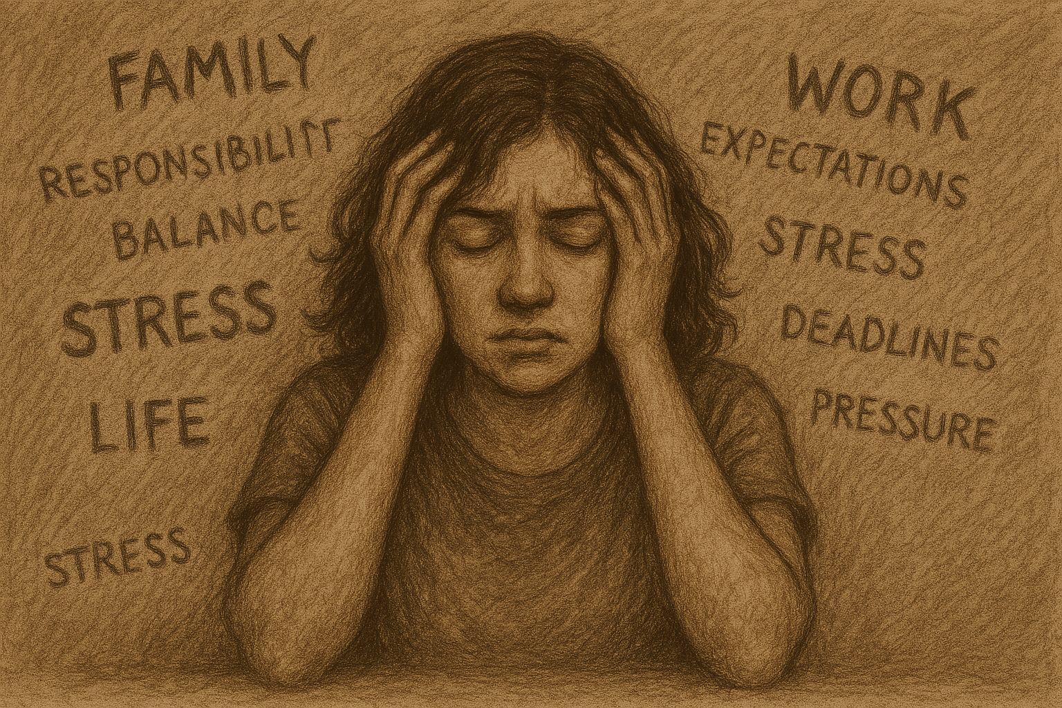
Figure 1: The Addiction Potential of ADHD Medications
Created by Sharleen Mtesa. This image shows the risk of addiction associated with ADHD medications when misused.

Psychostimulants like nicotine, cocaine, and amphetamines—think methamphetamine, MDMA, and even ADHD meds like Adderall are often seen as the bad guys in the world of substance use disorders. And they are, in many ways, wreaking havoc on both physical and mental health. But what’s even more concerning is how these drugs impact the brain’s ability to adapt and rewire itself. When the brain’s natural ability to learn and adjust gets disrupted, recovery becomes that much more challenging.

Therefore, researchers have turned their attention to metabotropic glutamate receptors (mGluRs)—crucial players in learning, memory, and brain plasticity. Emerging evidence suggests that mGluRs play a central role in how the brain rewires itself in response to repeated drug use [1]. And this opens the door to potential therapies that could help us manage, or even prevent, the long-term effects of psychostimulant use disorders (SUDs). 

The Highs and Lows of ADHD Meds: How Amphetamines Hack Your Brain

Let’s zoom in on one class of psychostimulants: amphetamines. You’ve probably heard of Adderall and Vyvanse, common medications used to treat ADHD. While they are effective tools for improving focus and attention, they come with a more complex story. Chemically, amphetamines are closely related to street drugs like meth and cocaine, which makes them especially powerful in the way they affect the brain’s reward system.

Not only do they boost dopamine and glutamate levels, but they also interact with Group I metabotropic glutamate receptors (mGluRs) [2] —specifically mGluR1 and mGluR5. According to the paper “A review on the role of metabotropic glutamate receptors in neuroplasticity following psychostimulant use disorder,” shows that these receptors become more active and start to reorganize in the brain [1]. And the medial prefrontal cortex (mPFC)? well this reorganization is especially important in the medial prefrontal cortex, which plays a key role in decision-making and cognitive functions. Changes in the mPFC can affect attention, impulse control, and overall thinking, helping explain why long-term stimulant use impacts these abilities.

So what’s the fallout from all this rewiring? When the mPFC increases its mGluR5 receptor activity, it makes the brain more likely to link the drug with pleasure, long after the first dose. This explains why people who misuse prescription stimulants can develop cravings and get hooked. The addictive potential is real, and understanding these effects is crucial, especially when it comes to medications.

ADHD Medications: Powerful Tools or Potential Pitfalls?

ADHD medications like Adderall, Vyvanse, and Ritalin are often life-changers for people struggling with focus, attention, and impulse control. These medications work by boosting levels of dopamine and norepinephrine [3] in the brain, allowing individuals to stay on task, manage their emotions, and excel in school or at work. For those with ADHD, these medications can be a game-changer, providing the structure and focus they need to thrive. But here’s the twist, when these same medications are misused, whether to stay awake, cram for exams, or chase a euphoric high, they hijack the brain’s reward system.

What Are Adderall and Vyvanse?

Adderall is a combination of two stimulant drugs: amphetamine and dextroamphetamine. By increasing the levels of dopamine and norepinephrine in the brain, Adderall helps improve focus, attention, and impulse control. The immediate-release version typically takes about 30-60 minutes to kick in and lasts around 4-6 hours, while the extended-release (Adderall XR) form provides longer-lasting effects of 10-12 hours.

Vyvanse, on the other hand, contains lisdexamfetamine, which is a prodrug. This means the body must metabolize it into its active form, primarily in the liver. It takes a bit longer to kick in, usually around 1-2 hours, but its effects can last up to 12-14 hours. Because Vyvanse is a prodrug, it has a smoother release, which might make its effects feel less intense compared to Adderall

The Dopamine Pathway: The Power and Pitfalls of ADHD Medications

One of the core reasons why ADHD medications like Adderall and Vyvanse are so effective is because they increase dopamine levels, especially in areas of the brain like the prefrontal cortex and striatum.

How it works:

Dopamine is released into the synapse. Under normal conditions, dopamine is reabsorbed by the neuron through a process called reuptake. But stimulants like Adderall and Ritalin block the dopamine transporter (DAT), preventing dopamine from being reabsorbed. This causes dopamine to accumulate in the synapse, enhancing focus, attention, and motivation.

Key Areas Affected:

  • Prefrontal Cortex: This region controls attention, decision-making, and impulse control.
  • Striatum: This area is involved in reward, motivation, and habit formation.

The Norepinephrine Pathway: Fueling Focus and Alertness

In addition to dopamine, these medications also boost norepinephrine, another neurotransmitter that’s important for alertness and focus.

How It Works:

Norepinephrine is released from nerve endings and binds to receptors that help increase alertness and focus. Stimulants like Adderall and Vyvanse block the norepinephrine transporter (NET), preventing norepinephrine from being reabsorbed. As a result, norepinephrine stays active in the synapse for longer, helping to maintain focus and attention.

Key Areas Affected:

  • Locus Coeruleus: This region regulates arousal, stress response, and alertness.


Figure 2: Stimulant Mechanisms of Action in ADHD Medications
This figure shows how ADHD medications like amphetamine (AMPH) and methylphenidate (MPH) increase dopamine (DA) and norepinephrine (NE) levels by affecting the dopamine transporter (DAT) and norepinephrine transporter (NET). AMPH and MPH promote the release of these neurotransmitters through DAT phosphorylation and reverse efflux, inhibiting reuptake and enhancing synaptic activity. [4]

Why This Can Lead to Addiction

While these medications are incredibly helpful for those with ADHD, they can also become addictive if misused. When Adderall or Vyvanse is taken for reasons other than prescribed—like to stay awake longer, study harder, or experience a euphoric high, the mesolimbic dopamine pathway, also known as the reward pathway, becomes activated. This pathway connects the ventral tegmental area (VTA) to the nucleus accumbens, creating powerful feelings of euphoria.

As a result, the brain starts to reinforce this behavior, making the individual crave more of the drug. Over time, this cycle of use and reinforcement increases the risk of addiction.

Figure 3: This figure highlights the brain areas impacted by ADHD, including the prefrontal cortex, basal ganglia, limbic system, and reticular activating system. Each area plays a key role in attention, behavior, emotional regulation, and impulse control. In ADHD, deficiencies in dopamine lead to symptoms such as inattention, impulsivity, emotional volatility, and hyperactivity. [5]

The Bitter Aftertaste of a Sweet Fix

So, here’s the bottom line: Adderall and Vyvanse are like superheroes for people with ADHD, helping them focus, stay on track, and crush their goals. But just like every superhero has a dark side, these medications can have unintended consequences when misused. By messing with the brain’s reward system and making it more likely to crave that dopamine rush, they can lead to some serious addiction risks. Understanding how these medications work and how they can rewire the brain will help us strike the right balance between maximizing their benefits and minimizing the risks. After all, the goal is to keep ADHD in check, not to let the medication take control of the brain’s spotlight.


Footnotes:

[1] Mozafari, R., Karimi-Haghighi, S., Fattahi, M., Kalivas, P., & Haghparast, A. (2023). A review on the role of metabotropic glutamate receptors in neuroplasticity following psychostimulant use disorder. Progress in neuro-psychopharmacology & biological psychiatry, 124, 110735. https://doi.org/10.1016/j.pnpbp.2023.110735

[2] Niswender, C. M., & Conn, P. J. (2010). Metabotropic glutamate receptors: physiology, pharmacology, and disease. Annual review of pharmacology and toxicology, 50, 295–322. https://doi.org/10.1146/annurev.pharmtox.011008.145533

[3] Del Campo, N., Chamberlain, S. R., Sahakian, B. J., & Robbins, T. W. (2011). The roles of dopamine and noradrenaline in the pathophysiology and treatment of attention-deficit/hyperactivity disorder. Biological psychiatry, 69(12), e145–e157. https://doi.org/10.1016/j.biopsych.2011.02.036

[4] Dutta, C. N., Christov-Moore, L., Ombao, H., & Douglas, P. K. (2022). Neuroprotection in late life attention-deficit/hyperactivity disorder: A review of pharmacotherapy and phenotype across the lifespan. Frontiers in Human Neuroscience, 16. https://doi.org/10.3389/fnhum.2022.938501

[5] Jie, T. S. (2021, July 16). ADHD From a Scientific Point-of-View. UnlockingADHD. https://www.unlockingadhd.com/adhd-from-a-scientific-point-of-view/


Why We Never Forget: How the Brain Builds Stressful Memories

Artstract Created by S. Mohamed

Have you ever noticed how the most stressful or traumatic moments in life tend to stick with you forever? Maybe it was a car accident, a time where you almost drowned, or a time when you were truly afraid. While it might feel like a harsh part of life, science shows there’s a biological reason why these memories are so vivid and lasting.

The paper highlights that stressful experiences initiate powerful molecular changes in the brain that help form long-lasting memories, especially through the activation of glucocorticoid and NMDA receptors, two key players in the memory-encoding process.[1], [2].

What’s Going On in the Brain?

Understanding the relationship between stress and memory starts with the hippocampus, specifically the dentate gyrus, a region critical for memory formation. When we experience stress, like public speaking or being stuck in traffic, the brain releases glucocorticoids (like cortisol). These hormones interact with glucocorticoid receptors (GRs) and NMDA receptors, triggering a powerful signaling cascade called the ERK-MAPK pathway [2], [4].

This pathway transmits signals into the nucleus of neurons, where it leads to a specific epigenetic modification of histone proteins: a dual mark called H3S10p-K14ac. This modification essentially “unlocks” sections of DNA so that crucial immediate early genes (IEGs) like c-Fos and Egr-1 can be transcribed into genes known to be essential for memory consolidation and synaptic plasticity [1],[4].

Figure 1. Psychological stress activates glucocorticoid and glutamate pathways that converge on histone modifications to promote stress-related gene transcription.

Why It Matters to You

This isn’t just fascinating neuroscience; it has real-world implications. The research helps explain why some individuals are more susceptible to PTSD and anxiety disorders after trauma. People with heightened GR/ERK-MAPK signaling might over-consolidate traumatic memories, while those with dysregulated GABA (a calming neurotransmitter) may lack the internal braking system to moderate stress responses [3].

Interestingly, the study also shows that this specific epigenetic switch—H3S10p-K14ac—isn’t used universally across the brain. While many brain regions show c-Fos and Egr-1 activation under stress, only the dentate gyrus requires this epigenetic tag for expression, indicating a uniquely guarded gateway to memory encoding in this area [1].

Another major revelation? Glucocorticoids don’t act alone. They rapidly enhance the ERK-MAPK pathway by physically interacting with phosphorylated ERK, accelerating the stress response and memory formation. This synergy happens within minutes—much faster than traditionally believed for steroid hormones [2].

What You Can Do About It

Thankfully, our brains aren’t helpless in the face of stress. GABA plays a protective role by maintaining inhibitory control in the hippocampus. Drugs like lorazepam (a benzodiazepine) can block stress-induced epigenetic responses, while anxiogenic compounds like FG7142 can amplify them. But you don’t need pharmaceuticals to influence this system.

Natural interventions, like long-term aerobic exercise, mindfulness, deep breathing, and quality sleep all help strengthen GABAergic tone and reduce stress-induced ERK signaling. Rats with access to running wheels showed less stress reactivity in the dentate gyrus, supporting the idea that movement can help buffer the brain from chronic stress [4].

This research doesn’t just explain how stress becomes a memory, it shines a light on how we might intervene. Targeting signaling proteins like MSK1 or transcription factors like Elk-1 could pave the way for new therapies that disrupt maladaptive stress memories while preserving helpful ones [2], [4].

Figure 2. Non-pharmaceutical approaches to anxiety include therapy, alternative treatments, and lifestyle changes that support mental wellness.

The Bigger Picture

We often think of memory as a passive thing events happen, and we either remember or forget. But the truth is, our brains are constantly evaluating which experiences deserve to be encoded. Stressful or emotional events? The brain gives those the red carpet treatment [1].

This molecular precision is part of our evolutionary design it helps us learn from danger. But sometimes, the system overreacts, reinforcing painful memories and fueling cycles of anxiety or trauma. Understanding how stress, hormones, and gene expression intersect gives us the power to reshape how we respond and remember [4].

A Better Ending: Hope for Healing and Resilience

What this research really highlights is that the brain is not static. Stressful experiences can leave deep biological marks, but those marks are not unchangeable. Through interventions like regular exercise, mindfulness, and even potential drug therapies targeting specific molecular pathways, we can change how our brains respond to and store stressful experiences.

In a time where anxiety and trauma are becoming increasingly common, especially among young people, it’s empowering to know that we can take an active role in our mental health. The more we understand about the biology of stress and memory, the better equipped we are to build resilience not just by avoiding stress, but by responding to it in healthier, more adaptive ways.

Stress may be unforgettable, but so is healing. And now, we have a clearer blueprint for how to get there.

References:

  1. Reul, J. M. H. M. (2014).
    Making memories of stressful events: A journey along epigenetic, gene transcription, and signaling pathways.
    Frontiers in Psychiatry, 5, 5.
    https://doi.org/10.3389/fpsyt.2014.00005
  2. Gutierrez-Mecinas, M., et al. (2011).
    Long-lasting behavioral responses to stress involve a direct interaction of glucocorticoid receptors with ERK1/2-MSK1-Elk-1 signaling.
    Proceedings of the National Academy of Sciences, 108(33), 13806–13811.
    https://doi.org/10.1073/pnas.1103214108
  3. McEwen, B. S. (2007).
    Physiology and neurobiology of stress and adaptation: Central role of the brain.
    Physiological Reviews, 87(3), 873–904.
    https://doi.org/10.1152/physrev.00041.2006

PTSD and C-PTSD: Memory Scars of Stress

Review of  “Making memories of stressful events: a journey along epigenetic, gene transcription, and signaling pathways” by Johannes M. H. M. ReulThe article by Reul explains how the brain forms and stores memories of stressful events. It takes a deeper look into the molecular parts of this process. He outlines how exposure to stress activates signaling pathways (particularly through glucocorticoid hormones and noradrenaline) that lead to epigenetic modifications and gene transcription changes in neurons, especially within the hippocampus and amygdala. These biological shifts affect how memories are formed, consolidated, and retained, which then often leaves lasting marks on an individual’s mental health. Reul has an important point in his paper that stress memory formation is not simply a switch that turns on or off, but a complex process that varies depending on the intensity and duration of the stressor, as well as individual genetic and environmental factors [1].

The Biology Behind PTSD and C-PTSD: Stress Leaves a Mark

Stress is something we all know, to varying degrees. A traffic jam, a missed deadline, or a tense conversation are all moments that come and go. But what happens when stress doesn’t just pass? What happens when it affects the biology of our brains? This is the core of Post-Traumatic Stress Disorder (PTSD) and Complex PTSD (C-PTSD), where memories of trauma don’t fade, but persist. They are vivid, intrusive, and disruptive [2].

CPSTD vs PTSD: definitions, similarities and differences - Priory
See the similarities and differences between CPTSD and PTSD here.


This matters because millions of people globally live with PTSD or C-PTSD, often silently [3]. Veterans, survivors of abuse, refugees, and even those exposed to chronic stress in childhood or adulthood carry the weight of painful memories that persist. These aren’t just psychological burdens. They are deeply biological and rooted in how our brains process and encode trauma at a molecular level.

Understanding this is crucial, because trauma isn’t a rare event. It’s a public health issue at this point in society. The science behind stress memory formation, like the work detailed by Johannes Reul, reveals not only the how behind trauma memory, but also the why (why some people recover while others remain stuck) [1].

This understanding is far from complete. PTSD and C-PTSD can be difficult to treat. Traditional talk therapy and medication can help, but not for everyone. The brain’s response to traumatic stress is very nuanced, involving cascades of molecular signals, epigenetic changes, and structural shifts in brain areas like the amygdala and hippocampus. For people with C-PTSD (often linked to prolonged, repeated trauma like childhood abuse rather than PTSD’s acute event), the challenge can be much more complex. Their brains don’t just react to a single traumatic event; they adapt to a world in which trauma is the norm [2].

Memory is Key

But the core issue lies in memory: why are these traumatic memories so persistent? Why do they resurface without warning? Why are they embedded so deeply in the fabric of who we are?

The insights from Reul’s research outline the journey from stress exposure to lasting memory through molecular biology. He reveals potential intervention points. For instance, if we can modulate how stress hormones affect gene transcription right after a traumatic event, could we prevent PTSD from developing? Could future therapies target epigenetic markers to “soften” traumatic memories without erasing them entirely [1]?

This research doesn’t just decode how trauma shapes memory. It also looks at a possible course toward healing. It tells us that trauma is not just stored in the psyche, but also in the synapses, genes, and proteins that shape our brain’s response to the world [4].

The Mind and Soul Foundation : PTSD
Here we can see the difference in memory consolidation between a “normal” brain and a brain experiencing trauma.

Societal and Future Impact

So what does this mean for us, for society, for the future? It means rethinking trauma care as not just a psychological issue, but a neurobiological one. It means investing in early intervention, especially for children and communities exposed to chronic stress. It raises powerful questions: Can we “edit” traumatic memories without compromising identity? What ethical challenges arise from altering memory? Would we one day vaccinate against PTSD?

And here’s the takeaway: Trauma is not just remembered- it is encoded. Understanding the biology behind PTSD and C-PTSD allows us to move beyond stigma and into a future where treatment is personalized and preventative. If the brain can change in response to trauma, it can also change in response to care.

Let’s keep asking questions. How do we turn understanding into healing? And let’s stay curious, because the science of memory isn’t just about the past, it’s about shaping the future.

Created by Rachel Cavaness, CHATGPT


[1] Reul, J. M. H. M. (2014). Making Memories of Stressful Events: A Journey Along Epigenetic, Gene Transcription, and Signaling Pathways. Frontiers in Psychiatry, 5. https://doi.org/10.3389/fpsyt.2014.00005

[2] Danfeng Li, Jiaxian Luo, Xingru Yan, & Yiming Liang. (2023). Complex Posttraumatic Stress Disorder (CPTSD) as an Independent Diagnosis: Differences in Hedonic and Eudaimonic Well-Being between CPTSD and PTSD. Healthcare, 11(1188), 1188. https://doi.org/10.3390/healthcare11081188

[3] Guideline Development Panel for the Treatment of PTSD in Adults, American Psychological Association. (2019). Summary of the clinical practice guideline for the treatment of posttraumatic stress disorder (PTSD) in adults. The American Psychologist, 74(5), 596–607. https://doi.org/10.1037/amp0000473

[4] Herman, J. (2012). CPTSD is a distinct entity: comment on Resick et al. (2012). Journal of Traumatic Stress, 25(3), 256–7; discussion on 260–3. https://doi.org/10.1002/jts.21697


Spam prevention powered by Akismet