Stress: The Memory That Sticks

Original Artstract by Meg Shercliffe

Original artstract by M. Shercliffe.

We’ve all experienced moments of intense stress that seem burned into our memories, whether it’s a near-miss car accident, a traumatic event, or even the anxiety of a final exam. But how does the brain cement these stressful experiences into long-term memory? The paper “Making Memories of Stressful Events: A Journey Along Epigenetic, Gene Transcription, and Signaling Pathways” [1] explores the molecular events behind this process, revealing a complex interaction between hormones, neurotransmitters, and epigenetic modifications. 

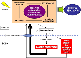
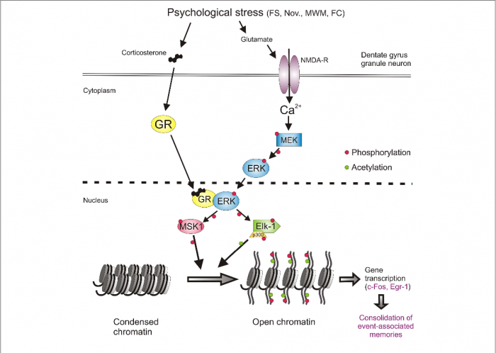
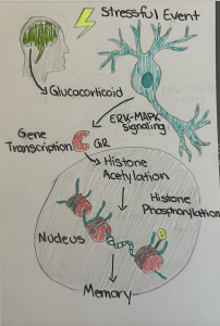
Understanding the relationship between stress and memory formation begins with the hippocampus, a brain region critical for memory formation. When faced with psychological stress (like a rat being forced to swim in a lab tank, or a human speaking in front of a large crowd), the hippocampus becomes activated, in turn activating a cascade of molecular events that help encode the experience. Neurons firing in the hippocampus are not the only factor, glucocorticoids (stress hormones like cortisol in humans, or corticosterone in rats) play a significant part.

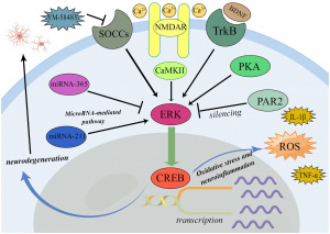
The paper highlights that these hormones don’t just passively float around, they actively shape how neurons respond to stress by interacting with glucocorticoid receptors (GRs). GRs act in combination with NMDA receptors, a glutamate receptor essential for learning and memory. Together, they trigger a signaling pathway involving ERK-MAPK, a series of proteins that relay stress signals into the nucleus of neurons.

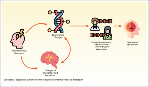
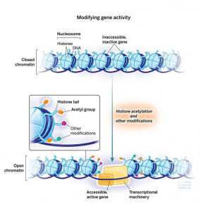
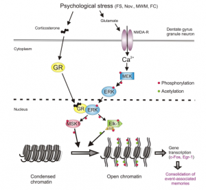
Inside the nucleus, this stress-induced signaling leads to epigenetic modifications: chemical tags on DNA and its associated histones that regulate gene expression without altering the genetic code itself. Specifically, stress causes two key changes on histone H3, and is demonstrated in the psychological stress pathway in Figure 1 below:

  1. Phosphorylation at serine 10 (S10)
  2. Acetylation at lysine 14 (K14)

This dual mark, H3S10p-K14ac, acts like a molecular switch, opening up tightly wound DNA so that genes can be transcribed. Which genes get turned on? Immediate-early genes (IEGs), like c-Fos and Egr-1, are crucial for synaptic plasticity and memory consolidation[1].

Figure 1. Psychological stress-activated signaling pathways in the dentate gyrus granule neurons. Psychological stress activates the GR and NMDAR/ERK/MAPK pathways [1].

However, this epigenetic mechanism isn’t universal across the brain. While many regions activate c-Fos and Egr-1 during stress, only in the dentate gyrus (a subregion of the hippocampus) does their expression require H3S10p-K14ac[1]. This suggests that dentate gyrus neurons keep these genes inactivated under normal conditions, only releasing them when stress demands it.

What’s novel about this research is that it illustrates how glucocorticoids and glutamate signaling collaborate. Traditionally, steroid hormones like glucocorticoids were thought to act slowly, binding to receptors in the nucleus and modulating gene expression over hours. However, this research suggests that GRs physically interact with phosphorylated ERK to increase the activity of downstream kinases like MSK1 and Elk-1[1].

This interaction happens within minutes of stress, allowing glucocorticoids to amplify glutamate-driven signals and accelerate epigenetic modifications. Thus, GRs serve to amplify the ERK-MAPK pathway, ensuring the stressful memory is highly encoded.  

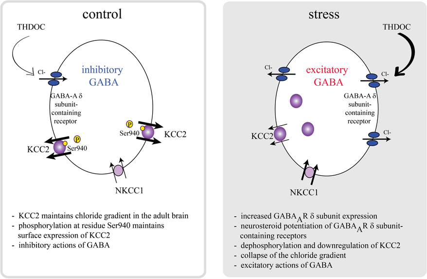
Thankfully, the brain doesn’t let stress run unchecked. GABA, the brain’s primary inhibitory neurotransmitter, modulates the dentate gyrus’s response to stress through a baseline level of inhibition. This finding is supported by studies of drug interactions:

  • Anxiolytics (like lorazepam, a benzodiazepine) suppress stress-induced H3S10p-K14ac and c-Fos activation.
  • Axiogenics (like FG7142, a GABA-A receptor antagonist) do the opposite, increasing epigenetic and gene responses.

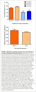
Even exercise plays a role. Long-term voluntary running in rats reduces anxiety-like behavior and dampens stress-induced ERK-MAPK signaling, likely by enhancing GABA synthesis[1]. This fits with real-world observations that exercise can help mitigate stress disorders[2].

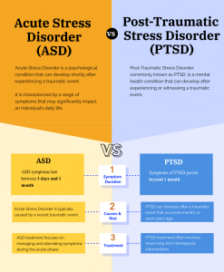
Understanding these mechanisms has real implications for stress-related disorders like PTSD and anxiety. In vulnerable individuals, the balance between stress-induced memory formation and adaptive coping may go awry.

  • Hyperactive GR/ERK-MAPK signaling: This might lead to the over-consolidation of traumatic memories (e.g. PTSD flashbacks).
  • Dysregulated GABA control: Could result in excessive fear responses or failure to dampen stress reactions[1].

This also opens doors for potential therapies. If researchers can pinpoint drugs that target these epigenetic or signaling pathways (like MSK1 or Elk-1) there’s potential to disrupt maladaptive stress memories without affecting useful ones.

This research paints a vivid picture of how stress etches itself into our brains. It’s not just about neurons firing, it’s a combination of hormones, neurotransmitters, and epigenetic marks. While evolution likely designed this system to help us remember threats, sometimes it overshoots, leaving us stuck in loops of anxiety or trauma. The good news? The brain is extremely plastic. Exercise, therapy, and even pharmacological interventions can adjust these pathways, offering hope for better stress resilience. 

Stressful events trigger glucocorticoids and glutamate signaling in the hippocampus, but the formation of long-term memories requires a precise epigenetic switch (H3S10p-K14ac) that activates memory-related genes. Therefore, understanding this mechanism reveals potential therapeutic targets for stress-related disorders like PTSD and anxiety.


References

[1] J. M. H. M. Reul, “Making Memories of Stressful Events: A Journey Along Epigenetic, Gene Transcription, and Signaling Pathways,” Front. Psychiatry, vol. 5, p. 5, Jan. 2014, doi: 10.3389/fpsyt.2014.00005.

[2] H. Alizadeh Pahlavani, “Possible role of exercise therapy on depression: Effector neurotransmitters as key players,” Behav. Brain Res., vol. 459, p. 114791, Feb. 2024, doi: 10.1016/j.bbr.2023.114791.

Stress Leaves a Mark – Literally: Why the Way We Remember Trauma Matters

We all experience stress. And we all remember those moments when life seemed overwhelming—finals week, heartbreak, a fender bender, or worse. These memories shape how we see the world and how we respond to future challenges. But have you ever wondered why some stressful experiences stay with us for years, while others fade quickly? Or why some people develop anxiety disorders or PTSD after trauma, while others don’t? Therefore, it’s essential that we understand not just that we remember stress—but how our brain encodes it, right down to the molecular level.

The Science of Stressful Memories: A Look Inside the Brain

In the article Making Memories of Stressful Events by Dr. Johannes Reul, a remarkable discovery is unpacked: when we undergo psychological stress, our brain physically changes to record that experience—starting with our DNA.

Here’s how it works: stress triggers the release of glucocorticoid hormones (like cortisol), which bind to glucocorticoid receptors (GRs) in the brain—especially in the hippocampus, the region responsible for memory. At the same time, glutamate activates NMDA receptors, kicking off a signaling cascade called the ERK-MAPK pathway. [1]

The magic happens when these two systems converge, forming a biological memory-making machine. This convergence leads to changes in the structure of histones, the proteins that package DNA. Specifically, a dual histone mark (H3S10 phosphorylation and K14 acetylation) appears, unlocking genes like c-Fos and Egr-1 which are crucial for memory consolidation. [1]

Figure 1 [2] Artstract by Ella Alsleben. Stressful experiences trigger glucocorticoid release and ERK-MAPK signaling in the brain, leading to epigenetic modifications like histone acetylation and phosphorylation in neurons. These molecular changes promote gene transcription that consolidates the memory of the event.

Why Should You Care

This research explains why some people develop disorders like PTSD after trauma—and others don’t. Only about 10–20% of people develop lasting mental health issues after a major stressor. This suggests that the way our brains process stress at the molecular level plays a major role in our mental resilience. [3]

Frontiers | The reciprocal regulation of stress hormones and GABAA receptors

Figure 1 [4]: Showing the difference between GABA in a normal functioning adult under non-stressful conditions and one under stressful conditions.

The study also revealed that GABA, the brain’s calming neurotransmitter, modulates this memory-encoding process (see Figure 1). More anxious individuals have lower GABAergic tone, which means their brains are more reactive to stress, forming stronger stress memories. Conversely, exercise was shown to increase GABA and reduce the brain’s stress response. [1]

So if you’ve ever wondered whether daily exercise can really help manage anxiety—the answer is, yes, even at the level of your DNA.

What Should You Take Away From This

This isn’t just a neuroscience geek-out—it’s a story about how our bodies remember, and how we can influence those memories.

If you’re a student, knowing that high anxiety makes stressful moments “stick” more might encourage you to seek out mental wellness tools before finals week (Figure 2).

If you’re managing anxiety, exercise isn’t just about physical health—it’s an epigenetic intervention.

Techniques to Reduce Stress and Anxiety

Figure 2 [5]: Wellness tools that can help relieve stress.

And if you’re someone who’s endured trauma, this science brings hope. Understanding the pathways that encode stress memories means we are one step closer to therapies that can help “unwrite” them.

Let’s Reframe the Conversation

Instead of viewing stress as something abstract or purely emotional, we can now see it as a physical imprint (Figure 3), a story our neurons etch into our DNA. And that story is shaped by biology, yes—but also by environment, habits, and resilience.

Understanding this empowers us to care for our mental health not just with willpower, but with scientific insight.

So the next time someone tells you that stress is “all in your head”—you can smile and say, “Yes, and it’s in my chromatin too.”

Brain Puzzles Images – Browse 161,646 Stock Photos, Vectors, and Video | Adobe Stock

Figure 3 [5]: Each story shapes our biology into the person we are today.

References

[1] Reul J. M. (2014). Making memories of stressful events: a journey along epigenetic, gene transcription, and signaling pathways. Frontiers in psychiatry, 5, 5. https://doi.org/10.3389/fpsyt.2014.00005

[2] Artstract by Ella Alsleben

[3] Schneiderman, N., Ironson, G., & Siegel, S. D. (2005). Stress and health: psychological, behavioral, and biological determinants. Annual review of clinical psychology, 1, 607–628. https://doi.org/10.1146/annurev.clinpsy.1.102803.144141

[4] Mody, I., & Maguire, J. (2012). The reciprocal regulation of stress hormones and Gabaa receptors. Frontiers in Cellular Neuroscience, 6. https://doi.org/10.3389/fncel.2012.00004

[5] Rebecca Valdez, M. (2024, June 10). Techniques to reduce stress and anxiety. Verywell Health. https://www.verywellhealth.com/how-to-reduce-stress-5207327

[6] Brain+puzzles images – browse 163,431 stock photos, vectors, and video. Adobe Stock. (n.d.). https://stock.adobe.com/search?k=brain%2Bpuzzles

I Can Only Stress the Importance

The article we have covered in a previous week, “Making Memories of Stressful Events: a journey along epigenetic, gene transcription, and signaling pathways” by Johannes M. H. M. Reul was an article about stressful events having a long lasting impact on both behavior and memories. Basically, we know already that memories are a feature of having a brain. However, due to this feature, between ten to twenty percent of human beings has developed some kind of stress disorder because of at least one traumatic events.The topic today is why people should care about this topic and what the people must know, so without further ado let’s get reading!


The article informs us of an experiment where they took lab mice through an experience where they put the mice in an enclosure which they flooded. Though, the article specifically focuses mainly on reactions between the initial and repeated attempts at this where the mice eventually began to connect the pieces together and began floating naturally. As a result of this connection, we can now classify here that traumatic moments can very much form memories.


Figure one, pictured above, from the article mentioned above is especially excellent at explaining this where it was needed (excellently timed, or in other words placed well). That piece is a bar chart expressing the response of these lab rats upon getting forced to swim for long periods of time. Although, one slight problem could be that it could feel a large bunch overwhelming if you’re not too familiar with the applied terms of figure one in the Neuroscience field. This was an excellent piece to me for it is maximized simplicity because, for clear reasons, that kind of thing strongly helps. The figure may also benefit people uninvolved in Neuroscience as well because figure one uses so much brief, yet descriptive, labeling, and I find that effective myself in general because it’s easy on the eyes to track or logicate.


Now, at this point, one, such as yourself, may wonder why people really should care about all the above information. Well, let’s answer with essential basics to answer ourselves by quickly asking ourselves something simpler first; what really is an anxiety disorder? Well, the answer is absolutely nothing short of a true tragedy. According to Cleveland Clinic, an anxiety disorder is known as “a group of mental health conditions that cause fear, dread and other symptoms that are out of proportion to the situation” (Cleveland Clinic 2025). Considering that we know about these symptoms as a consequence, it’s no shock to pretty much anyone that such a scenario can turn serious fast. Let us put this into perspective with another topic.


In my class, I personally examined the various animal model types used in studies of stress. Surprisingly, I learned from authors at Science Direct of various vertebrate animals, such as animals we often think of as pets (pigs, dogs, rabbits, and so on) used to model the effects stressful events have on the heart muscle (Nicola Maggio and Menahem Segal, 2019). It is nothing short of incredible that we have the option to use inhuman specimens to reduce the need for real human beings in experiments that would be way too dangerous for us all, but I insist that we must subscribe to love and ethics when we do so.


Works Cited:
1) “Making Memories of Stressful Events: a journey along epigenetic, gene transcription, and signaling pathways” by Johannes M. H. M. Reul

2) Cleveland Clinic 2025 on anxiety disorders

3) “Stress, Corticosterone, and Hippocampal Plasticity” by Nicola Maggio and Menahem Segal, 2019

Trauma’s Code: Stress Signals and the Epigenetic Blueprint

Artsract by J. Copiskey

Trauma’s Code: Stress Signals and The Epigenetic Blueprint

Stress is a universal human experience, yet its impact on the brain often goes unnoticed. It leaves a lasting molecular and cellular footprint, shaping emotions, behaviors, and memories. By unraveling the science behind stress responses, we can better understand how stress rewires the brain and explore the critical roles of neurotransmitters and epigenetic changes in trauma recovery.

Understanding PTSD, Anxiety, and Stress

Anxiety and stress disorders are widely prevalent among individuals with PTSD, producing emotional and physiological responses that affect overall well-being. The interactions between stress and emotional or cognitive processing can reveal how such mechanisms contribute to the development and persistence of PTSD and stress.

Fig. 1

The process of converting DNA instructions into RNA is fundamental for driving cellular responses; in this case of stress,  glucocorticoid hormones are involved, which play a pivotal role in regulating the brain’s stress response and the functionality of the hippocampus, which is a region essential for memory and emotional regulation.

GABAergic neurons are crucial, which regulate brain excitability, and the limbic brain structures, including the amygdala and hippocampus, as the emotional core of the brain.

Finally, the role of vital signaling pathways, such as ERK-MAPK, and transcription factors like CREB, emerges as essential contributors to the molecular changes induced by stress. As shown in Fig.2.

Fig.2

What is an Epigenetic Pathway?

Epigenetic pathways are powerful mechanisms that regulate how stress impacts the brain, without altering the underlying DNA sequence. These processes involve modifications to histones, which are proteins that DNA wraps around. As well as chemical additions like methylation or acetylation, which influence whether specific genes are “switched on” or “off.”

In the context of stress and PTSD, epigenetic changes can increase  or suppress gene expression, particularly in areas like the hippocampus and amygdala. For example, stress-induced histone modifications, shown in Fig. 3 such as serine10 phosphorylation and lysine14 acetylation, play a role in activating immediate early genes (IEGs) critical for neural adaptation and behavioral responses. [4]

These epigenetic marks have effects across signaling pathways like ERK-MAPK,  reshaping how the brain responds to fear and anxiety over time. Such changes can be enduring, encoding trauma and contributing to long-term vulnerability to PTSD,  The dynamics between stress and the epigenetic marks emphasize the brain’s remarkable adaptability and synaptic plasticity. [5]

Fig. 3


Acute Stress vs Chronic Stress

  • Acute stress can sometimes be beneficial, as it primes the brain for quick responses. However, chronic stress is a significant risk factor for anxiety disorders.
  • Chronic stress or PTSD induce changes in the amygdala, PFC, and hippocampus which contribute to heightened anxiety, impaired emotional regulation, and difficulty distinguishing between real and perceived threats.


Fig.4

Acute Stress

Acute stress is short-term and typically arises in response to immediate threats or challenges, such as a sudden deadline or a near-miss car accident.

The brain’s amygdala detects the threat and signals the hypothalamus, which activates the sympathetic nervous system. This triggers the “fight-or-flight” response, releasing stress hormones like adrenaline and cortisol.

Effects on the Brain:

  • Enhancement of glutamate release in the PFC, improving focus and decision-making temporarily.
  • Activates the hippocampus, aiding memory formation related to the stressful event.
  • Once the stressor is resolved, the parasympathetic nervous system restores balance, reducing cortisol levels.[1]

Chronic Stress

Chronic stress occurs when stressors persist over time, such as ongoing financial difficulties or a toxic work environment.

This causes prolonged activation of the hypothalamic-pituitary-adrenal (HPA) axis , which then leads to sustained cortisol release.

Affects on the Brain

  • Hippocampus: Chronic cortisol exposure can cause atrophy of hippocampal neurons, impairing memory and learning. Chronic cortisol causes dendritic shrinkage and spine loss, impairing episodic memory and spatial navigation.
  • Prefrontal Cortex: Reduced activity in the PFC weakens decision-making and emotional regulation. Dendrites in the medial PFC retract, impairing memory and emotional resilience, while the orbitofrontal cortex (OFC) shows dendritic expansion associated with hypervigilance.
  • Amygdala: Becomes hyperactive, heightening fear and anxiety responses. Acute stress increases dendritic growth temporarily, but chronic stress leads to more enduring expansions in basolateral amygdala (BLA) dendrites, heightening anxiety and fear responses.
  • Neuroplasticity: Chronic stress disrupts synaptic plasticity, leading to long-term changes in brain circuits associated with anxiety and depression. [2]


Acute Stress Chronic Stress
Short-term HPA axis activation. Prolonged HPA axis activation and dysregulation.
Boosts PFC activity for focused thinking. Impairs PFC function, weakening decision-making and emotional regulation.
Enhances hippocampal memory encoding. Causes hippocampal atrophy and reduced neurogenesis.
Balanced glutamate levels support plasticity. Excess glutamate triggers excitotoxicity.
Parasympathetic system restores balance. Persistent cortisol alters brain circuits.

Stress and Structural Changes in The Brain

Anxiety and stress disorders are common among individuals with PTSD, producing emotional and physiological responses that can affect overall well-being. Critical aspects include gene transcription, the process of converting DNA instructions into RNA to drive cellular responses, and the role of glucocorticoid hormones, which influence the brain’s ability to respond to stress. The adrenal cortex produces corticosterone, which is a vital glucocorticoid hormone involved in the body’s stress response. As shown in Fig. 5

Corticosterone plays a central role in regulating metabolism, immune response, and the HPA axis. When the body encounters stress, the adrenal cortex releases corticosterone into the bloodstream to help maintain homeostasis. This hormone influences the energy balance by increasing glucose availability, ensuring that the body has enough resources to respond to the stressor.

Fig. 5

Additionally, the hippocampus, is highlighted alongside GABAergic neurons that manage brain excitability through the inhibitory neurotransmitter GABA. Furthermore, limbic brain structures such as the amygdala and hippocampus, as well as critical signaling pathways like ERK-MAPK and transcription factors such as CREB, play a significant role in the stress-induced molecular changes.

Neurotransmitters Involved in Stress

  • Glutamate: As the brain’s primary excitatory neurotransmitter, glutamate enables rapid communication between neurons. Acute stress uses glutamate effectively to enhance cognitive performance, but chronic stress floods the brain, causing excitotoxicity and disrupting synaptic plasticity. Glutamate is critical for associative learning  and emotional processing under stress. Dysregulated glutamate signaling contributes to distortions in how PTSD patients process information. [3]
  • GABA (Gamma-Aminobutyric Acid): The brain’s primary inhibitory neurotransmitter, GABA helps counterbalance glutamate’s excitatory effects, calming hyperactive neurons. Chronic stress reduces GABA function, heightening anxiety and emotional reactivity.
  • Dopamine: Acute stress briefly boosts dopamine, aiding motivation and focus. Chronic stress depletes dopamine, contributing to loss of pleasure and depression.
  • Serotonin: Regulates mood and emotional processing. Stress can diminish serotonin levels, increasing vulnerability to anxiety and depression.
  • Noradrenaline (Norepinephrine): Heightens alertness and attention during acute stress but can lead to heightened vigilance and overreaction under chronic stress.

These neurotransmitters form the biochemical foundation for how the brain processes and adapts to stress, playing a critical role in shaping behaviors and emotional outcomes in PTSD. As seen in Fig. 6  Optimal conditions versus stressful conditions in a neurotransmitter.

Fig. 6


Why This Matters

Stress and PTSD  leave lasting imprints on the brain, from changes in neurotransmitter function to epigenetic marks that shape how we process fear, memory, and resilience. We can understand the brain’s plasticity allows it to adapt to stress, but also how chronic stress can disrupt this balance, leading to vulnerabilities like anxiety, depression, and PTSD.

Understanding the molecular and cellular effects of stress gives us more than knowledge, it gives the potential future of treatment. By targeting pathways like ERK-MAPK or leveraging therapeutic potential in neurotransmitters such as GABA and glutamate, science paves the way for innovative treatments that go beyond symptom management to address the root causes of trauma.

REFRENCES

Gudsnuk, K., & Champagne, F. A. (2012). Epigenetic influence of stress and the social environment. ILAR journal53(3-4), 279–288. https://doi.org/10.1093/ilar.53.3-4.279

Howie, H., Rijal, C. M., & Ressler, K. J. (2019). A review of epigenetic contributions 
to post-traumatic stress disorder
. Dialogues in clinical neuroscience21(4), 417–428. https://doi.org/10.31887/DCNS.2019.21.4/kressler

JM;, R. (n.d.). Making memories of stressful events: A journey along epigenetic, gene transcription, and signaling pathways. Frontiers in psychiatry. https://pubmed.ncbi.nlm.nih.gov/24478733/

Martin, E. I., Ressler, K. J., Binder, E., & Nemeroff, C. B. (2009). The neurobiology of anxiety disorders: brain imaging, genetics, and psychoneuroendocrinology. The Psychiatric clinics of North America32(3), 549–575. https://doi.org/10.1016/j.psc.2009.05.004

FOOTNOTES

[1] JM;, R. (n.d.). Making memories of stressful events: A journey along epigenetic, gene transcription, and signaling pathways. Frontiers in psychiatry. https://pubmed.ncbi.nlm.nih.gov/24478733/


[2] JM;, R. (n.d.). Making memories of stressful events: A journey along epigenetic, gene transcription, and signaling pathways. Frontiers in psychiatry. https://pubmed.ncbi.nlm.nih.gov/24478733/


[3] Martin, E. I., Ressler, K. J., Binder, E., & Nemeroff, C. B. (2009). The neurobiology of anxiety disorders: brain imaging, genetics, and psychoneuroendocrinology. The Psychiatric clinics of North America32(3), 549–575. https://doi.org/10.1016/j.psc.2009.05.004

[4] Howie, H., Rijal, C. M., & Ressler, K. J. (2019). A review of epigenetic contributions 
to post-traumatic stress disorder
. Dialogues in clinical neuroscience21(4), 417–428. https://doi.org/10.31887/DCNS.2019.21.4/kressler

[5] Gudsnuk, K., & Champagne, F. A. (2012). Epigenetic influence of stress and the social environment. ILAR journal53(3-4), 279–288. https://doi.org/10.1093/ilar.53.3-4.279


The Stress-Memory Puzzle: Why Studying the Human Brain Isn’t So Simple

previewer
An abstract by Venesa Angau


Stress Enhances Some Memories—but Not Always

It’s well-known that stressful events are often remembered more vividly than neutral ones. From an evolutionary standpoint, this makes sense: remembering where the lion chased you could save your life. But the biology behind this is anything but simple.

Reul (2014) explains that strong psychological stressors initiate a cascade of molecular changes in the hippocampus and amygdala, key regions involved in learning, memory, and emotion. In animal studies, stress-related memory formation relies heavily on glucocorticoid hormones like cortisol (or corticosterone in rodents), which are released during stress and bind to glucocorticoid receptors (GRs) in neurons​ [1].

Simulating Stress in a Lab is Tricky

In animal studies, stress can be induced through tests like the forced swim test or Morris water maze, which reliably elevate glucocorticoids and activate memory formation pathways. But in humans, ethically inducing high levels of stress is much harder—researchers can’t just  dunk people in cold water or give them electric shocks.

This means that lab-based stress tests (like giving a surprise math quiz or asking participants to speak in front of a crowd) often fall short of triggering the deep biological mechanisms that actual trauma or survival-related stress would.

Not Everyone Reacts to Stress the Same Way

One of the biggest obstacles is individual variability. Some people under stress develop strong, clear memories. Others forget details or block the event entirely. This is partly explained by variations in anxiety levels and GABAergic control, as Reul’s study highlights​ [1].

In the hippocampus, a person’s anxiety level influences how their neurons respond to stress at the molecular level. Higher anxiety tends to amplify stress responses—via both hormone release and epigenetic changes—making certain memories stronger or more “sticky” [2].

Epigenetics: The Memory Code That Keeps Changing

MAPK Erk pathway - Cusabio
Figure 1. As shown in this figure is the MAPK-ERK Signaling Pathway [3].

Stress doesn’t just flip a switch in your brain; it rewires it—literally. Reul and colleagues found that stress activates the ERK-MAPK signaling pathway, which leads to a specific modification on histone proteins: H3S10 phosphorylation and K14 acetylation (H3S10p-K14ac). This chromatin remodeling opens up DNA for transcription of “immediate-early genes” like c-Fos and Egr-1, which are critical for long-term memory consolidation​.

But this change doesn’t happen uniformly across the brain. These epigenetic marks are highly specific to areas like the dentate gyrus in the hippocampus, and are influenced by prior experiences, local interneuron activity (especially GABAergic tone), and afferent input from emotional centers like the amygdala [4].

We Still Don’t Know the Full Picture

Despite decades of research, we still can’t fully predict who will remember a traumatic event clearly, who will develop PTSD, and who will walk away largely unscathed. Reul’s research shows that the consolidation of stress-related memories depends on an intricate interaction between hormones, neurotransmitters, genetics, and environmental context.

The bottom line? Studying stress and memory is hard because the human brain is built for complexity. It’s not just about remembering what happened—it’s about how your biology, mood, history, and genes all converge in that moment.

And in the end, understanding that convergence might just be the key to treating stress-related disorders like PTSD and anxiety.

Resources

[1] Reul, J. M. H. M. (2014). Making memories of stressful events: A journey along epigenetic, gene transcription, and signaling pathways. Frontiers in Psychiatry, 5, Article 5. https://doi.org/10.3389/fpsyt.2014.00005

[2] Robinson, O. J., Vytal, K., Cornwell, B. R., & Grillon, C. (2013b). The impact of anxiety upon cognition: Perspectives from human threat of shock studies. Frontiers in Human Neuroscience, 7(203). https://doi.org/10.3389/fnhum.2013.00203

[3] MAPK Erk pathway – Cusabio. (n.d.). Retrieved from www.cusabio.com website: https://www.cusabio.com/pathway/MAPK-Erk-pathway.html

[4] Kandler, C. (2021, June 22). Epigenetic Regulation – an overview | ScienceDirect Topics. Retrieved from www.sciencedirect.com website: https://www.sciencedirect.com/topics/psychology/epigenetic-regulation


Reinforcing Anxiety Through Memories


Having an anxiety-ridden experience can cement into our memories, so it lives on for years afterward. For some people, there may only be a couple memories full of anxiety we can remember; however, for people with anxiety disorders, these memories become frequent. Typically, these memories can come up in daily lives and lead to people believing the worst-case scenario will occur. Anxiety allows us to adapt, but in anxiety disorders, it can impair daily functioning and turn daily activities or interactions into threats.

We will look at different anxiety disorders and symptoms, the science behind anxiety memories, as well as how anxiety can impair memory formation in daily life.

Anxiety Disorders

Some physical symptoms of anxiety include:

  • Restlessness or feeling on edge
  • Sleep difficulties
  • Increased heartrate
  • Sweating
  • Shaking
  • Hyperventilating

Psychological symptoms:

  • Trouble concentrating
  • Racing thoughts
  • Difficulty controlling worry
  • Avoiding anxiety-triggering interactions/places/situations

Some common anxiety disorders include Generalized Anxiety Disorder (GAD), Social Anxiety Disorder, Post Traumatic Stress Disorder (PTSD), Phobias, and other anxiety disorders.

Biological Pathways

Glucocorticoids are released in response to stress, they’re stress hormones. They go inside cells and activate Glucocorticoid Receptors (GR). These receptors can phosphorylate, or activate, ERK. ERK can further activate MSK and Elk-1, which will lead to the unraveling of DNA, the chromatin. [1]

Figure 1: Biological pathway for Anxiety Memory Formation [2]
This unraveling of the chromatin will allow for gene transcription to take place. [3] Gene transcription allows for memory formation, so the stress induced activity becomes ingrained in the mind.

This is especially prevalent in PTSD. [4] Trauma memories can disrupt daily functioning and become invasive. People at higher base-line levels of anxiety may be more prone to these anxiety-induced memories to form. [5]

Daily Memories and Anxiety

Anxiety in the classroom can distract students from learning and forming memories about the lesson content. People with Generalized Anxiety Disorder (GAD), characterized by anxiety or worry around multiple areas of life and a difficulty in controlling it, have reported difficulties with memory recall. [6]

It’s hypothesized that anxiety consumes so much mental energy, that little mental resources are left for memory encryption, storage, and recall. Working memory, which is short term memory of items told to you, such as someone’s phone number, is thought to be impaired in GAD and PTSD. [7]

While anxiety-memories seem to increase and have a strong long-lasting impact in anxiety disorders, creating and remembering memories for during daily life may be impaired while the person is experiencing anxiety.

Conclusion

Anxiety disorders have a variety of physical and psychological symptoms, but it does impact daily life. In the brain, we see stress hormones released during anxiety that leads to the unraveling of DNA. This unraveling of DNA leads to memory formation. These strong anxiety memories create a cycle of anxiety as the person will go to worst-case scenarios for daily life.


References

[1-5] Reul, J. M. H., (2014). Making memories of stressful events: a journey along epigenetic, gene transcription, and signaling pathways. Frontiers in Psychiatry, 5(5). doi: 10.3389/fpsyt.2014.00005

[6, 7] Gkintoni, E., & Ortiz, P. S. (2023). Neuropsychology of Generalized Anxiety Disorder in Clinical Setting: A Systematic Evaluation. Healthcare (Basel, Switzerland)11(17), 2446. https://doi.org/10.3390/healthcare11172446

Exercise is Medicine: How Moving Your Body Calms Your Mind

Abstract created by G. Sparks

Memories play a huge role in our everyday lives. We’re constantly forming them, whether we realize it or not, and they help us navigate the world around us. Built from our experiences and environments, memories allow us to adapt, grow, and prepare for similar situations in the future.1 But not all memories are created equal. Our brains tend to hold onto stressful or traumatic events more strongly, creating lasting imprints that can shape how we think and feel. Over time, these intense memories can contribute to the development of anxiety in certain situations. In more severe cases, they may even lead to conditions like post-traumatic stress disorder (PTSD).

PTSD can seriously impact a person’s quality of life, often bringing nightmares, flashbacks, negative thought patterns, mood swings, and other overwhelming symptoms.1 Many people coping with PTSD or chronic anxiety turn to therapy or medication for relief and those treatments can be incredibly helpful. But what if there were also a natural way to help ease those symptoms? What if something as simple as movement, like exercise that could play a role in healing the brain?

Recent research has been exploring exactly that. Studies show that exercise can act as a form of medicine, helping the brain and body gradually shift out of survival mode and into a healthier, more balanced state.

Understanding the Science Behind Anxiety

When it comes to the science of anxiety, researchers have proposed many pathways to explain how stress impacts the brain. One particularly interesting pathway involves gene transcription and how it contributes to the consolidation of event-associated memories, especially those tied to stressful or traumatic experiences.1

Two key players in this process are corticosterone (a stress hormone) and glutamate (a neurotransmitter). During a stressful event, these molecules work together to set off a cascade of biological events that ultimately encode the memory of that event into long-term storage.

Here’s how it happens: Glutamate binds to NMDA receptors located on dentate gyrus granule neurons in the hippocampus. This binding allows calcium ions to flow into the cell, initiating a signaling pathway that activates MEK, which in turn activates ERK through phosphorylation. ERK then travels into the nucleus of the neuron.

At the same time, corticosterone enters the cell and binds to glucocorticoid receptors (GR), which also move into the nucleus. Once inside, ERK and GR interact, allowing pERK1/2 to phosphorylate two important molecules: MSK1/2 and Elk-1. These molecules then go on to modify the H3 histone by phosphorylating and acetylating it.

This modification causes the chromatin, which was previously tightly wound around the histones and inaccessible, to loosen. This unwinding opens up the DNA, allowing gene transcription to occur. The result is the consolidation of the memory associated with that stressful event.

A visual representation of this pathway is shown in Figure 1 below. For a more detailed explanation of the molecular cascade and its components, refer to the full article.


Figure 1. A diagram that illustrates the pathway of corticosterone and glutamate signaling leading to the consolidation of event associated memories. 1

Exercise and Its Role in Reducing Stress and Anxiety

Traditionally, medication and therapy have been the two primary treatments for individuals dealing with anxiety. While both can be highly effective, medications sometimes come with side effects or interactions, especially for people managing other conditions like ADHD.1 In recent years, however, there’s been a growing interest in the therapeutic potential of exercise as a powerful, natural intervention for anxiety.

Research has shown that regular aerobic exercise can reduce the reactivity of both the sympathetic nervous system and the hypothalamic-pituitary-adrenal (HPA) axis which are two key systems involved in the body’s stress response.2 The HPA axis plays a central role in regulating cortisol levels, and exercise has been found to induce long-term adaptations that help moderate stress reactivity and reduce anxiety symptoms.

Exercise also influences several important neurotransmitter systems. In animal studies, physical activity has been linked to increased levels of monoamines such as serotonin, dopamine, and norepinephrine which are chemicals that are commonly targeted by antidepressant medications.2 These changes can produce an antidepressant-like effect and help improve mood stability.

Another important system affected by exercise is the opioid system. Physical activity triggers the release of beta-endorphins, which are natural painkillers and mood enhancers.2 This release is believed to contribute to the well-known “runner’s high” and can significantly lower perceived levels of stress and anxiety.

Additionally, exercise has been shown to increase levels of brain-derived neurotrophic factor (BDNF) which is a key neurotrophin that supports the growth and resilience of neurons.2 Elevated BDNF levels are associated with better emotional regulation, enhanced cognitive function, and improved mental health outcomes.

Together, these biological effects suggest that exercise doesn’t just distract from anxiety, but that it may actually reshape the brain’s response to stress in a meaningful and lasting way.

Here is another link to an article that takes an in depth look on the effects of exercise and physical activity on anxiety.

Final Thoughts

Anxiety is a complex condition that’s closely tied to how our brain stores and responds to stressful events. In some cases, these memories are helpful since they prepare us to react and adapt when similar situations arise in the future. But when the stress is too intense or traumatic, those memories can become harmful. In some individuals, this can lead to the development of PTSD, a condition that often reduces quality of life through persistent fear, anxiety, and emotional distress.

While medications and therapy are effective for many people, they aren’t perfect solutions and can come with complications or limitations. Fortunately, a growing body of research points to exercise as a promising complementary approach. Though it can’t erase trauma, regular physical activity may be a reliable and accessible way to improve mood and reduce the physical and emotional burden of stress.

Whether it’s going for a walk, lifting weights, movement has the power to heal. Exercise truly is a form of medicine, and it might just be one of the most effective tools we have for supporting mental health.

References

(1)      Reul, J. M. H. M. Making Memories of Stressful Events: A Journey along Epigenetic, Gene Transcription, and Signaling Pathways. Frontiers in Psychiatry. 2014. https://doi.org/10.3389/fpsyt.2014.00005.

(2)      Anderson, E.; Shivakumar, G. Effects of Exercise and Physical Activity on Anxiety. Front Psychiatry 2013, 4 (APR). https://doi.org/10.3389/fpsyt.2013.00027.


Addiction Rewires Your Brain: Can We Rewire It Back?

Artstract created S. Mohamed

Addiction is not a choice. It’s a complex brain disease. And for millions of people struggling with psychostimulant use—cocaine, meth, nicotine, and amphetamines—it can feel like there’s no way out. While we know these drugs hijack the brain’s reward system, we’re still uncovering how they reshape it at the molecular level. Until now, there hasn’t been a strong therapeutic target to reverse these brain changes and truly help people recover. But new research offers a powerful insight: a group of brain receptors called metabotropic glutamate receptors (mGluRs) might be the key to healing the brain after drug abuse.

Therefore, understanding how these receptors work could change the way we treat—and talk about—addiction. Let’s unpack how psychostimulants mess with our brain chemistry and why that makes addiction less of a choice and more of a brain health issue.

The Reward Circuit and Dopamine: Why Drugs Feel Good

Dopamine is the brain’s “feel-good” chemical. It spikes when you experience something pleasurable like eating chocolate, getting a compliment, scrolling TikTok at 1 a.m. It also spikes when someone uses a drug. And that’s where the problem starts.

Dopamine plays a huge role in the brain’s reward system, which includes areas like the ventral tegmental area (VTA), nucleus accumbens (NAc), and prefrontal cortex (PFC). When dopamine floods this system, the brain takes note: “This felt good. Let’s do it again.” Over time, your brain builds tolerance, needing more and more of the drug just to feel normal.

But there’s another layer: the brain learns to expect dopamine in certain contexts maybe it’s a particular room, a group of friends, or even a certain time of day. These environmental triggers can increase tolerance or make someone more likely to relapse. And when the drug is taken outside of that context, the body isn’t ready leading to accidental overdose[2].

Figure[1]. This figure illustrates the brain’s reward circuit connecting the prefrontal cortex, nucleus accumbens, and ventral tegmental area via dopamine signaling and how its dysregulation contributes to addiction.

Glutamate: Reinforcing the Habit

While dopamine makes drugs feel good, glutamate helps the brain remember how good it felt.

Glutamate is the brain’s main excitatory neurotransmitter. It’s essential for learning, memory, and synaptic plasticity a fancy term for how the brain strengthens or weakens connections between neurons. Drugs like cocaine, nicotine, and amphetamine increase glutamate levels and hijack these learning processes.

They do this by activating metabotropic glutamate receptors (mGluRs)—especially Group I receptors like mGluR1 and mGluR5. These receptors become overexpressed or relocated to different parts of neurons. For example, mGluR5 increases in the prefrontal cortex, while mGluR1 spreads into extrasynaptic sites, both changes linked to long-term neuroplasticity in addiction[1].

The result? The brain gets really good at learning how to crave drugs and not so great at resisting them.

Figure [2].  Brain regions and synaptic pathways involved in addiction, showing how Group I, II, and III mGluRs differentially modulate glutamatergic and dopaminergic signaling.

Double Trouble: Rewired and Unbalanced

Normally, your prefrontal cortex helps with decision-making and impulse control. But repeated drug use weakens this system. The brain keeps getting messages like this drug is great from the reward pathway, while the logical part of your brain can’t shut it down.

So… Can the Brain Ever Go Back?

The scary thing about addiction is how good the brain becomes at remembering drugs. But the hopeful part? The brain is also capable of change. Researchers have found that those same metabotropic glutamate receptors (mGluRs) that help reinforce drug memories might also hold the key to reversing them. By targeting these receptors, it may be possible to rebalance the brain’s glutamate system, reduce cravings, and actually “unlearn” the cycle of addiction.

Here’s what’s exciting:

  • Blocking mGluR5 reduces drug-seeking and relapse in animal studies.

  • Activating mGluR2/3 helps calm excessive glutamate and lowers craving.

  • These strategies work across multiple substances like cocaine, meth, nicotine, and more.

And most importantly, these treatments don’t just patch over symptoms. They work at the cellular level, where addiction takes root. They aim to restore balance in how the brain learns, remembers, and responds to reward.

Addiction Is Brain Health

So yeah, drug use and addiction is scary. It rewires how the brain is rewarded, how the brain learns, and how much impulse control a person has. But the biggest takeaway here is that addiction is not just bad choices—it’s bad brain chemistry.

Understanding the science helps us reframe the narrative. Addiction isn’t a personality flaw. It’s a brain disorder that deserves real treatment and real compassion. The more we know about glutamate, dopamine, and mGluRs, the closer we get to treating addiction like the health condition it truly is.

References:

  1. Mozafari, R., et al. (2023). A review on the role of metabotropic glutamate receptors in neuroplasticity following psychostimulant use disorder. Progress in Neuropsychopharmacology & Biological Psychiatry.
  2. Siegel, S. (2001). Drug tolerance, cue exposure, and overdose. Addiction Research & Theory, 9(5), 411-419.
  3. Kalivas, P. W., & Volkow, N. D. (2005). The neural basis of addiction: A pathology of motivation and choice. Am J Psychiatry, 162(8), 1403-1413.

Learning How Anxiety is Disrupted Learning

Artstract created C. Geraci

Do you know someone with an anxiety disorder or do you, yourself, have one? Have you ever wondered what is going awry in the brain to make that happen? To understand this, we need to establish a baseline understanding on how memory formation and consolidation, which is the process of creating a permanent long-term memory, occur.


The Anatomy of the Limbic System

The limbic system is the primary part of the brain involved in memory formation and consolidation, and the primary structures within it are the basal ganglia, amygdala, and hippocampus. Each of these structures is portrayed in Figure 1 along with the types of memory they encode, but below each memory type is defined along with its corresponding brain structure.1

  • Basal ganglia:  procedural memory, which remembers how we certain perform tasks.
  • Hippocampus: declarative or explicit memory, which involves remembering things like names, facts, etc.
  • Amygdala: implicit & procedural memory, with implicit memory involving remembrance of things not outrightly spelled out but more likely felt, like emotions.1


Figure 1. Illustrates the limbic system’s structures and what types of memory each is involved in creating.1


The Hippocampal Formation, Specifically the Dentate Gyrus

Figure 2. Illustrates the anatomy of the dentate gyrus in the hippocampus formation.2


The dentate gyrus (DG) is part of something called hippocampal formation, which also encompasses the hippocampus (see Figure 2). It is made of multiple types of cells and is involved in the formation of episodic memory and exploration of new environments.

The three types of cells are dentate granule cells, which are excitatory and use glutamate as their primary neurotransmitter, inhibitory dentate pyramidal basket cells, and excitatory, unmyelinated granule cells called mossy fibers. Mossy fibers synapse onto the CA3 region of the hippocampus, which contains neurons that can then send excitatory signals to the CA1 region of the hippocampus.  However, all these play a role in either exciting or inhibiting hippocampal neuron firing.2


The Pathway of Memory Formation, Learning, & Memory Consolidation

Figure 3. Illustrates the different lobes of the cerebral cortex and the cerebellum’s role in memory formation.1


Also involved in memory formation are the cerebral cortex and cerebellum, and for information on the cerebellum, click here. The cerebral cortex is where certain memories are stored after the hippocampus has consolidated them, with each lobe corresponding to a different type of memory stored there, as can be seen in Figure 3 above.1

Compiling all the above information, we can create the below pathway that illustrates how memory formation and learning occurs:

  1. Learning begins when sensory signals are transcribed in the cerebral cortex
  2. These signals are transmitted to either the hippocampus, amygdala, and/or dentate gyrus. If to the amygdala or dentate gyrus, they send signals the correlate to how and if the memory is processed by the hippocampus
  3. If the signal is strong or repeated, a long-term memory is consolidated by the hippocampus
  4. The memory is wired back to the cerebral cortex for storage.3

And when you think about it, when you are able to recall how to do something or a fact, that is the outward sign that you’ve accurately consolidated a memory. Aka, you’ve learned something!


A Pathway to Anxiety

But in anxiety disorders, such as PTSD or generalized anxiety disorder, some part of this memory formation, learning, and/or consolidation process is disrupted. One study investigated why this is and discovered elevated levels of a dual histone mark called H3S10p-K14ac. This histone mark was found on the promoter regions of the fos and egr1 genes. Essentially, one part of these genes was phosphorylated while another part was acetylated, and what this caused was increased transcription of the fos and egr1 genes, leading to increased anxiety.4


Interestingly, the genes with the H3S10p-K14ac dual histone mark were only present in dentate neurons, and when GABAergic inhibitory signals were administered to the DG to block H3S10p-K14ac histone marks from building, symptoms of anxiety decreased. As established earlier, the DG plays a role in telling the hippocampus whether to consolidate a memory or not, so the fact that inhibition of block H3S10p-K14ac histone marks in the DG led to decreased anxiety tells us that something anxiety disorders disrupt the flow of information from the DG to the hippocampus. This makes it so memories cannot be correctly stored in the cerebral cortex, perhaps making them more present in a person’s mind, therefore causing them to feel anxiety in situations where if their memory had been correctly consolidated, they would not be making anxious associations with previously psychologically stressful experiences.4 For more information on how the dual histone H3S10p-K14ac mark forms following psychologically stressful events, click here.


Conclusion

Overall, there is a typical pathway by which learning and memory formation occur within the brain, with the limbic system and the cerebral cortex playing a large role in securely storing memories so that we can recall them at appropriate times. But anxiety disorders involve a disruption in this pathway, allowing memories and their associated emotions, like fear, to be at the forefront of someone’s mind at times they wouldn’t normally. Therefore, further research should be done to study what factors are disrupted in each anxiety disorder so that this information can be used to target the root biochemical cause of each disorder.


Footnotes:

1https://www.youtube.com/watch?v=yepwx67_UkM

2Shahid, Shahab. “Dentate gyrus.” Kenhub. 2023. https://www.kenhub.com/en/library/anatomy/dentate-gyrus#

3https://www.youtube.com/watch?v=4Hm08ksPtMo

4Reul, M.H.M. Johannes. “Making memories of stressful events: a journey along epigenetic, gene transcription, and signaling pathways.” Frontiers in Psychiatry, vol. 5, no. 5, 2014, doi: 10.3389/fpsyt.2014.00005

Childhood Trauma & Memory

Traumatic experiences in childhood have a more significant impact on the brain than acute trauma in adulthood. Let’s look into the field of developmental trauma, and one of the ways this is so impactful… memory.

Developmental Trauma

The field of developmental trauma is a new interdisciplinary area of study exploring how traumatic events during childhood affect an individual’s brain and nervous system [1]. 

Developmental trauma is the early, persistent, repeated attacks on healthy development. This includes abuse, neglect, maltreatment, disrupted attachments, exposure to violence, or distress not typical for a child. This type of trauma at such a critical period of development has been found to have a profound impact on the general health and well-being of individuals well into adulthood [2]. The impacts on the mind, brain, and body are not simply outgrown. Childhood trauma causes chronic activation of the stress response system, dysregulating the nervous system, and leading to an imbalanced fight or flight response [1]. This impacts relationships, resilience, and overall worldview. And causes affective, physiological, cognitive, behavioral, self, and relational symptoms [2].

The ACE Study

The first study looking into this correlation was the Adverse Childhood Experiences (ACE) study by the CDC and Kaiser Permanente published in 1998 [3]. This survey asks a series of questions meant to measure how many traumatic events happened in an individual’s childhood. Each question answered ‘yes,’ is one point. Significant evidence supports that increased ACE scores are correlated with decreased overall mental and physical health and well-being. For example, chronic illness, risk-taking behaviors, and suicidality all significantly increase as ACE scores increase. Additionally, individuals with higher ACE scores have a significantly greater chance of dealing with the United States’ top ten leading causes of death [3].

Memory

Artstract created by Hadlie Dahlseid.

One reason trauma is so impactful on anxiety is due to the learning and memory mechanisms of the brain. During a stressful event, glucocorticoid levels are increased, causing stronger associations with the event, and a stronger and longer-lasting memory. Stressors concurrently activate glucocorticoid receptors and NMDA receptors in the dentate gyrus of the hippocampus. When NMDA receptor and glucocorticoid receptor activity converge, histone phosphorylation is impacted. This ultimately leads to gene transcription related to memory consolidation of stressful events [4].

NMDA Receptors

Signaling pathway after activation of glucocorticoid and NMDA receptors [4].
When glutamate binds to a NMDA receptor, an ERK-MAPK signaling pathway is triggered. This is a key pathway involved in learning and memory [4].

Glucocorticoid Receptors

With the release of glucocorticoids, the glucocorticoid receptor interacts with ERK, facilitating the phosphorylation of downstream nuclear kinases, specifically Elk and MSK of the ERK/MAPK pathway. The glucocorticoid receptor acts like a scaffold in this process.

Histones

Next, the glucocorticoid receptor phosphorylates and acetylates histone H3, forming the H3S10p-K14ac mark. These histone modifications are epigenetic mechanisms that lead to altering chromatin structure, promoting the transcription of genes. This results in the entry of immediate-early genes (IEGs) which are essential for memory formation associated with the stressful event [4].  

Dealing with Trauma

So now what do we do with all this information? Trauma-informed care is an approach to social services that recognizes and responds to effects of trauma, while creating a safe space that reduces the risk of re-traumatization [5]. This includes practices to regulate the nervous system, therapies and medications such as those used to treat other anxiety disorders, and understanding the impacts of trauma on the brain and nervous system. Recognizing that trauma is something that happened to someone, not something wrong with someone, is vital. Society needs to shift from asking “what’s wrong with you?” to “what happened to you?”. We can start doing this by understanding the science behind childhood trauma.

References

[1] van der Kolk, B. (2014). The Body Keeps the Score: Brain, Mind, and Body in the Healing of Trauma. Penguin Books. 

[2] Perry, B. D., & Winfrey, O. (2021). What happened to you?: Conversations on trauma, resilience, and healing. Flatiron Books.

[3] Felitti, V. J., Anda, R. F., Nordenberg, D., Williamson, D. F., Spitz, A. M., Edwards, V., Koss, M. P., & Marks, J. S. (1998). Relationship of Childhood Abuse and Household Dysfunction to Many of the Leading Causes of Death in Adults: The Adverse Childhood Experiences (ACE) Study. American Journal of Preventive Medicine, 14(4), 245–258. doi:10.1016/S0749-3797(98)00017-8.

[4] Reul, J. M. (2014). Making memories of stressful events: A journey along epigenetic, gene transcription, and signaling pathways. Frontiers in Psychiatry, 5. https://doi.org/10.3389/fpsyt.2014.00005 

[5] What is trauma-informed care?. University at Buffalo School of Social Work – University at Buffalo. (2024, May 29). 

Spam prevention powered by Akismet